
Part 1 covered terminology, aetiology, classification and the gray zone of penile size. This part deals with the ground on which actual decisions are made: whether to operate, and if so when and how.
Indications — what the guidelines do not say
One thing should be made clear at the outset. There is no international guideline that defines the indication or the timing of surgery for concealed penis.
In the EAU/ESPU Paediatric Urology guideline, concealed penis is not a chapter of its own; it appears in narrative form inside "Phimosis and other abnormalities of the penile skin." The text distinguishes the types, notes that they must be differentiated from micropenis and aphallia, and mentions that buried penis due to abnormal prepubic fat distribution may resolve with growth or weight loss. There is exactly one graded recommendation, and it concerns not the indication for surgery but a contraindication to circumcision — do not perform simple circumcision when phimosis is associated with another penile anomaly such as buried penis (strong recommendation), because the foreskin may be needed for reconstruction. The AUA has no guideline on paediatric concealed penis.
The indications used in practice are therefore extracted retrospectively from published series: difficulty with hygiene, balanoposthitis, voiding disturbance (spraying, urine trapped in the prepuce, difficulty holding the penis), and psychological distress over appearance.
Pooling the reported frequencies, symptomatic patients make up around 40% at most, voiding symptoms around 35%, and inflammation including urinary tract infection around 25%. In a series of 35 boys with concealed penis after circumcision, 43% underwent surgery for symptomatic penile adhesion and 8% for balanitis.
A point of honesty is needed here. In the survey of parents of surgical patients that I am currently conducting, the most common reason for seeking care was not a symptom but concern about the size and appearance of the penis. Voiding and inflammatory complaints were reported less often. The results will be reported separately, but the direction is consistent with clinical experience. It is why the indication cannot be discussed in functional terms alone — and why reimbursement disputes are not easily settled.
Timing — a debate without consensus
Setting aside cases where fat is the only issue and pubertal growth can be expected to help, opinion is divided on the right time to operate.
Shapiro argued for early surgery: boys with a buried penis are aware of it before puberty, and early correction benefits psychological development. In Herndon et al.'s long-term follow-up, parental satisfaction was consistently higher when surgery had been done in infancy than in adolescence. Ferro and Philip recommend correction as soon as the diagnosis is made, to resolve both dysuria and the cosmetic problem.
By contrast, the parents surveyed by Shenoy preferred an age at which the problem could be discussed with the child — before secondary school.
I too have been surveying parents of surgical patients about the appropriateness of timing. But such surveys have a structural limitation: the respondents are almost all parents, so they cannot speak for the patient's own satisfaction or discomfort. Given the growing weight placed on the patient's own autonomy over surgery in the hypospadias literature, concealed penis cannot avoid the same question. Surgery in early childhood is the parents' decision, and it is the parents who then judge the result.
With that in mind, my current practice runs along two lines. Where the anatomical problem is clear — an inner-dartos abnormality or penoscrotal fusion — I recommend early correction, before toilet training. Where the anatomy is not severe and improvement can be expected with penile growth, I set the target after puberty. The reason for leaning toward early correction is wound healing. After puberty, androgen-driven erections put repeated tension on the suture line, sebaceous secretion and pubic hair make wound care harder, and thicker, more vascular tissue produces more oedema and more prominent scarring. When the tissues are small, the extent of dissection needed for the same correction is also smaller. This is my clinical standard, not an evidence-based recommendation, and the autonomy question raised above remains open within it.
Technique — common principles and their limits
A search of the last twenty years alone turns up more than thirty described techniques. Incisions and suture patterns vary, but four elements are emphasised across almost all of them.
- Radical dissection of the abnormal fibrous band and connective tissue
- Local skin flaps to cover deficient penile skin
- Fixation sutures between skin and corpora
- Relief of the phimotic ring
To these, lipectomy or liposuction of suprapubic fat and correction of penoscrotal fusion are sometimes added.
Hadidi's approach uses a ventral midline incision from the tip of the prepuce to the penoscrotal junction in all grades, with the content tailored by grade: the fundiform and suspensory ligaments are divided from Grade II, and excess fat is excised in Grade III. The tunica albuginea is fixed to the periosteum of the symphysis, the excess inner prepuce is excised, and the result is finished as a circumcised penis.
What is striking is how little the technique itself seems to change long-term outcome. Frenkl et al. concluded that the number of fixation sutures did not affect recurrence; Herndon reported that the procedure used did not significantly alter long-term results. In Hadidi's 61 patients, 5 in Grades II and III (9%) required reoperation for late partial retraction. If refining the technique does not change the result, what separates good outcomes from poor ones is more likely who was operated on — a question of indication.
How the technique came to change
From fellowship until 2017 I used the relatively minimal technique described by Sugita et al. (2010, J Urol): spatulation of the preputial opening with rotation of dorsal skin to the ventral side.
Problems emerged with longer follow-up. Some patients held their exposure well, but in a fair number the shape achieved at surgery had, a year later, reverted to a state where the pubic fat had to be pressed down for the penis to appear. Cases were seen with barely the glans exposed and poor overall exposure.
So I reviewed what parents were dissatisfied with after surgery. The complaints fell into three groups.
- The penis is still small after surgery
- As the swelling settles, the skin looks redundant
- Exposure is still poor after surgery
The response to each was different.
For the first, the conclusion was "secure penile size first, then decide about surgery." Size cannot be changed by surgery. These patients are typically somewhat small with abundant surrounding fat and no major morphological problem. The approach shifted to watching growth through puberty — or, where the penis is markedly small, securing size first with hormonal treatment — and only then reassessing exposure and deciding on surgery. This connects with the third problem as well: once size is secured, the same technique yields a different exposure.
The literature supports this. Hadidi stated explicitly that stretched penile length did not change after surgery in his 61 patients. Further, in a case followed for ten years the flaccid glans:shaft ratio remained 1:1 and the penis appeared shorter than normal, and the five patients followed beyond five years all appeared shorter than expected. Length gained intraoperatively by dissection and traction largely disappears in the flaccid state. To recommend surgery to a patient whose main complaint is size is to promise what cannot be delivered.
Second, redundant skin reflects the character of the tissue and has to be accepted to a degree. Some lymphoedema follows almost every concealed-penis repair, and as it settles the skin appears to sit in excess. As discussed in Part 1, the dartos in these patients has reduced and fragmented elastin, poor compliance, and probably poor lymphatic drainage; add surgical dissection and oedema forms easily and lasts. Oedema is a problem in itself, but more so because it changes the shape of the penis — the contour is blunted while it is swollen, and the skin looks redundant once it has settled. The two parental complaints, "still small after surgery" and "there is leftover skin," may well share a single root.
I do not, however, regard this as permanent. My observation is that it improves over time as the penis grows and lymphatic channels re-open. In particular, skin that looks redundant is drawn out and smoothed naturally as the penis grows — what appears to be excess now becomes the skin that covers the growing shaft. Ferro had already raised, from the surgical side, the concern that deep dissection could cause lymphatic stasis; the histological findings and the operative observation point the same way. This is a hypothesis rather than a verified mechanism. For now, the best course is to explain this expected course before surgery.
Third, poor exposure was the only complaint that surgical refinement could address. In retrospect, the Sugita technique had overlooked two things: removal of the fibrofatty tissue and correction of penoscrotal fusion. Widening the preputial opening and rotating skin left the structures tethering the penis inward untouched. The technique was changed to deal with both in a single operation.
Current technique
The principle is that excision of the fibrofatty tissue is the main manoeuvre and the penile skin is handled as little as possible. The approach is from the scrotal side, and no preputial skin is excised.
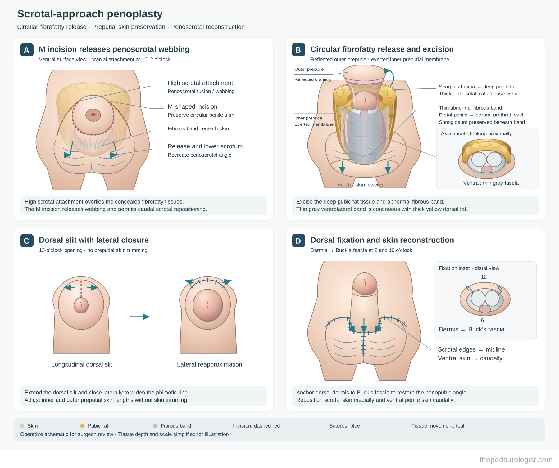
A. M-shaped penoscrotal incision. The incision follows the line of fusion between scrotum and penis in an M shape. It releases the webbing and at the same time creates room to reposition the scrotum caudally. The circular skin around the penis is preserved intact.
B. Release and excision of the circular fibrofatty tissue. With the outer skin reflected cranially and the inner prepuce exposed, the fibrofatty tissue encircling the penis is dissected. Ventrally it appears as a thin grey fibrous band, but dorsolaterally it becomes continuous with thick adipose tissue, and above it leads into Scarpa's fascia and the deep pubic fat. Following this continuity, the excision effectively includes a lipectomy of the deep pubic fat. Ventrally the corpus spongiosum lies directly beneath and is preserved during dissection. This step alone secures much of the penile exposure.
C. Dorsal slit with lateral closure. A longitudinal incision at the 12 o'clock position is extended and then closed laterally to widen the phimotic ring. No skin is excised; only the relative lengths of inner and outer prepuce are adjusted. Because the prepuce is preserved, tissue remains available should reconstruction ever be needed.
D. Dorsal fixation and skin reconstruction. The dorsal dermis is anchored to Buck's fascia at the 2 and 10 o'clock positions to establish the penopubic angle. The scrotal skin is then brought to the midline and the ventral penile skin repositioned caudally to reconstruct the penoscrotal angle.
Summarised in one line, the technique is radical soft-tissue excision with minimal skin manipulation — and it addresses every anatomical contributor — fibrofatty tissue, penoscrotal fusion, the phimotic ring and skin fixation — in a single operation.
Should circumcision be performed?
On the basis that a long inner prepuce was a constant finding in all 61 of his patients, Hadidi holds that circumcision should be performed in every case to excise the excess inner mucosa. Summerton et al., on the other hand, reported that redundant inner prepuce is not present in buried penis, and Redman observed no related structural abnormality in his cases. The literature contains both the claim that skin is redundant and the claim that it is deficient.
My own experience has led me to see this from a slightly different angle. When I used the Sugita technique, skin was left over at the end of the operation and a substantial amount of inner prepuce had to be trimmed away. The literature repeatedly reports that skin is deficient in concealed penis, yet here it was in excess and being excised. I found this odd.
After changing to the current technique, the redundant skin disappeared even though no skin was excised. Cases even appeared where the skin looked deficient. The interpretation converges on one explanation: the earlier technique did not fully correct the anatomical abnormality, and skin draped over uncorrected structures ended up redundant. Once the fibrofatty tissue and the penoscrotal fusion are properly released, that skin returns to where it belongs.
So my answer to whether circumcision should be performed is "it happens by itself." When the anatomical correction is complete, the skin settles correctly over the exposed penis and the result is the same as a circumcised penis. There is no need to decide in advance whether to excise the inner prepuce.
What remains
To summarise: definition and classification remain ambiguous, and studies of surgical indication and long-term outcome are lacking. More than thirty techniques have been described, but the evidence that any of them changes long-term outcome is weak. And counselling proceeds with the problems surgery can fix mixed together with those it cannot.
In an editorial comment on three papers about buried penis, Frank asked a provocative question — "Why do our adult urologist colleagues not see this condition in the older patient?" It may correct itself at puberty; adults may be diagnosed instead with a congenitally short penis or with a hidden penis due to fat; or patients may adapt to the condition and simply not seek advice.
We do not yet have the data to answer it. Until we do, I think the best a surgeon can do is to draw the indication narrowly and to say beforehand what surgery cannot fix. Satisfaction after this operation depends less on the refinement of the technique than on the setting of the indication and the explanation given before it.
References
- Casale AJ, Beck SD, Cain MP, Adams MC, Rink RC. Concealed penis in childhood: a spectrum of etiology and treatment. J Urol. 1999;162(3 Pt 2):1165-1168.
- Herndon CDA, Casale AJ, Cain MP, Rink RC. Long-term outcome of the surgical treatment of concealed penis. J Urol. 2003;170(4 Pt 2):1695-1697.
- Frenkl TL, Agarwal S, Caldamone AA. Results of a simplified technique for buried penis repair. J Urol. 2004;171(2 Pt 1):826-828.
- Sugita Y, Tanikaze S, Yoshino K, Yamamichi F. Severe chordee correction and concealed penis repair. J Urol. 2009;182(4 Suppl):1734-1737.
- Hadidi AT. Buried penis: classification surgical approach. J Pediatr Surg. 2014;49(2):374-379.
- Kim SW. Concealed penis from the perspective of pediatric urology. Urology Digest. 2021;2(2). (in Korean)
- EAU Guidelines on Paediatric Urology. Phimosis and other abnormalities of the penile skin. European Association of Urology, 2026.