
Part 2 covered why the staged Fowler–Stephens procedure came into wide use for the high intra-abdominal testis whose vessels are too short. Yet results of the same two-stage operation differ considerably between centres. In a recent study pooling 469 two-stage Fowler–Stephens procedures from six UK centres, atrophy was 22% overall but ranged from 11% to 37% between centres (Mohamed et al., 2025). Case mix, technique, follow-up and the way atrophy was judged may all have contributed. One factor examined repeatedly over the past decade is whether the gubernaculum is divided or preserved at the second stage.
A disclosure first: I have not yet performed this technique. This article works through the anatomy and the literature to answer for myself whether the testis really comes down with the gubernaculum preserved, and it keeps what the literature shows separate from my own interpretation. What is "preserved" here is the gubernacular tissue and the connecting vessels within it; it does not mean that every original attachment is left fixed in place.
Blood supply of the testis — arteries and veins
Arteries
The arterial supply of the testis is usually described as three systems, although their number and development vary. The testicular artery arises from the aorta just below the renal artery and descends retroperitoneally. The deferential artery arises from the internal iliac system (mostly the inferior vesical artery) and runs with the vas. The cremasteric artery branches from the inferior epigastric artery, itself a branch of the external iliac, near the internal ring and enters the cord with the cremaster. The three anastomose close to the testis, particularly around the tail of the epididymis and the lower pole (Benzi et al., 2022).
Their calibres differ. In 120 adult spermatic cords measured at the external ring, the testicular artery averaged 1.0 mm, the vasal artery 0.6 mm and the cremasteric artery 0.5 mm, and in 57.5% the testicular artery alone was at least as wide as the other two combined (Raman & Goldstein, 2004). The testicular artery is the relatively largest of the three — and it is precisely the vessel that Fowler–Stephens gives up.
Fetal anatomical data from normal descent show that the arrangement changes with testicular position. In 32 fetuses (64 testes), 71.9% had three arteries, 23.4% only two (testicular and deferential) and 4.7% four, and every testis with only two arteries was intra-abdominal (Sampaio et al., 1999; summarised in Benzi et al., 2022). The higher the testis, the shorter the testicular artery and the longer the deferential and cremasteric arteries, the latter reaching the lower pole where the gubernaculum attaches. Because these data come from normal fetal descent, they should not be read as the vascular distribution of the undescended testis in children. The gubernaculum carries the cremaster and its vessels.
Veins
The testicular vein, formed from the pampiniform plexus, drains into the IVC on the right and the left renal vein on the left. There are also deferential and cremasteric veins. The three systems connect at the lower pole and communicate through gubernacular veins with the posterior scrotal veins (Mirilas & Mentessidou, 2012). The same paper explains that, in varicocele surgery, once the internal spermatic and cremasteric veins have been ligated, ligating the gubernacular veins as well leaves venous return dependent on the deferential veins. As an anatomical paper on varicocelectomy, it invites thought about venous preservation in Fowler–Stephens, but direct clinical evidence for that is lacking.
The cremasteric artery is a named artery arising from the inferior epigastric artery. "Gubernacular vessels", by contrast, refers to the vessels of the gubernaculum and its surrounding tissue, veins included, so the two terms are not exact synonyms. A separate "gubernacular artery" is not usually named in descriptions of the normally descended testis. In this article, "cremasteric / gubernacular vessels" means the connecting vascular network preserved in the gubernacular region. It has also been suggested that vessels from tissue around the distal attachment contribute (Yamataka et al., 2003, as cited by Ellis et al., 2014).
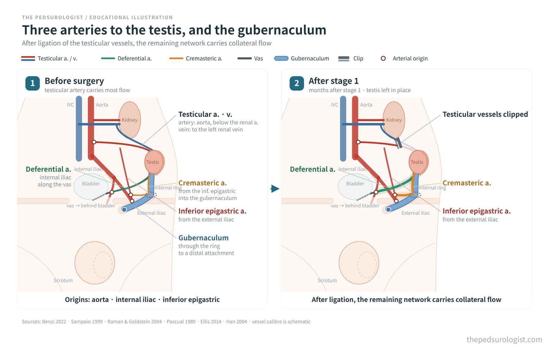
What remains after the testicular vessels are ligated
The rationale of the two-stage operation is that collaterals follow if time is allowed after ligation. Animal work first showed this. In rats, testicular blood flow fell by about 80% one hour after ligation of the testicular vessels and was back to baseline 30 days later (Pascual et al., 1989). When the vessels were ligated at 10 days of age, flow in adulthood was 18% lower, but testicular weight and spermatogenesis were normal (Pascual et al., 1990).
Direct human data on which vessels take over are scarce. The most direct are photographs taken at a gubernaculum-sparing second stage. In a surgical series of 25 patients and 28 testes, intraoperative photographs from selected patients showed well-developed cremasteric vessels running along the gubernaculum and anastomoses with the vasal vessels and the ligated testicular artery stump. The two systems were joined by several arching vessels just proximal to the epididymis; in some, a fine plexus replaced the larger arches. Calibre was not measured before and after ligation, so this shows a prominent collateral network at the second stage rather than proof that ligation enlarged it.
Clinically, every testis in the series survived (mean follow-up 3.3 years, size assessed by examination, a small non-comparative series), and one testis with congenitally absent vas was brought down on the gubernacular vessels alone after division of the testicular vessels (Ellis et al., 2014). Whether these collaterals are acquired after ligation, the authors wrote, would require comparing the vascular supply in single-stage and two-stage Fowler–Stephens. Another series observed that 10 of 25 testes had moved to a lower position during the six months after the first stage (Robertson et al., 2007).
In short, two collateral systems remain after the first stage: the deferential system along the vas and the cremasteric / gubernacular system entering along the gubernaculum from the internal ring. The former is preserved in any second stage. Whether the latter is preserved or lost depends on how the gubernaculum is handled at the second stage.
Where the second stage diverges
The traditional second stage divides the gubernaculum to free the testis, leaves it on a broad strip of peritoneum around the vas, and brings it to the scrotum through a new route medial to the inferior epigastric vessels (the Prentiss manoeuvre). This is shorter than the original path through the internal ring and canal — one study put the gain at about 1.5 cm (Abbasion et al., 2024). The testis then depends mainly on the deferential system, and it has been suggested that angulation along this route can interrupt the collaterals between the gubernaculum and the deferential artery (Ellis et al., 2014).
The gubernaculum-sparing approach typically brings the testis down on its gubernaculum through the internal ring and canal — the path it should have taken. The most detailed description runs as follows (Zhou et al., 2022). The peritoneum between the vas and vessels and the testis is incised, and a broad sheet of peritoneum including the vas, testis, gubernaculum and the connecting vessels between them is preserved and dissected free. The testicular vessels are divided between the clips placed at the first stage, and dissection is carried further along the vas to lift a wide peritoneal triangle containing the collaterals. Through a small scrotal incision, a laparoscopic dissector is passed up to the internal ring to create a tunnel, and a clamp passed along it grasps the testis at the gubernaculum and draws it into the scrotum. If tension remains, more peritoneum over the vas is freed.
Since it was first described (Wang & Shaul, 2004), the principle has been applied in several ways: an entirely laparoscopic second stage (Robertson et al., 2007; Braga et al., 2019), a laparoscopic-assisted second stage (Mahomed et al., 2012) or an open inguinal second stage (Zhou et al., 2022). It has been reported in single-stage Fowler–Stephens (Naycı et al., 2017), and a randomised trial applied it to conventional orchiopexy without division of the testicular vessels (Abbasion et al., 2024).
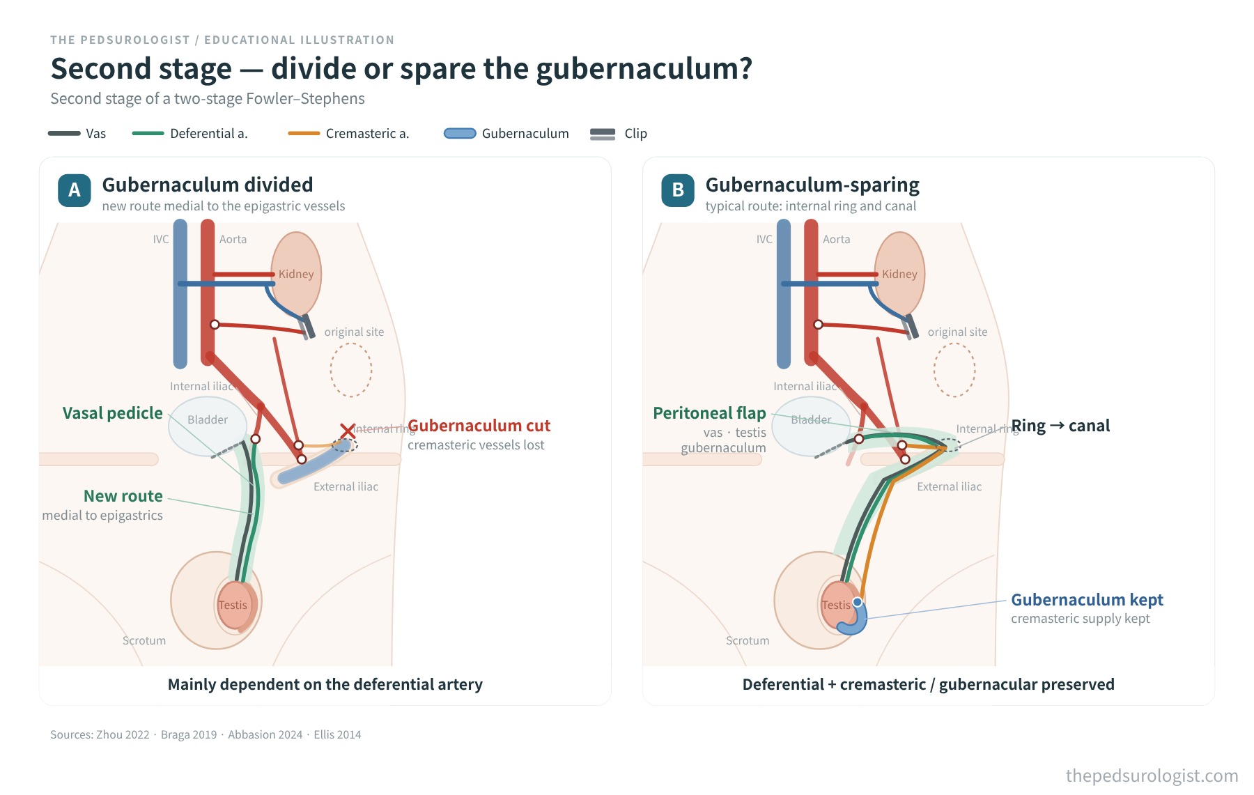
How the testis comes down with the gubernaculum preserved
A natural question arises on first meeting this technique. Undescended testis is often explained by an abnormal distal attachment of the gubernaculum. As discussed in Part 1, in the series of Han et al. the distal gubernacular attachment was abnormal in 93% of undescended testes, and of 142 impalpable abdominal testes it lay around the inguinal ring in 58% (83) and on the lateral or upper scrotum in 25% (36) (Han et al., 2004). Does it make sense to bring the testis down while preserving the very structure blamed for arrested descent? That observation, however, records the site of attachment; it does not show why a particular dissection works.
What the literature shows
The gubernaculum usually lies distal to the testis and is not described as the main structure holding the testis up at the second stage, although this depends on attachment site, length and route and is hard to generalise. A series of 205 gubernaculum-sparing second stages named the length of the vas as the main factor limiting final position and reported tension-free scrotal placement in 95.6% with adequate dissection (Zhou et al., 2022). The authors of the randomised trial likewise noted that excising the gubernaculum does not consistently lengthen the cord (Abbasion et al., 2024).
That is as far as the literature goes: the gubernaculum and its connecting vessels are preserved while the vas and surrounding peritoneum are dissected enough to bring the testis down (Zhou et al., 2022), and the distal attachment, gubernacular vessels and testis are moved together through the internal ring (Braga et al., 2018, meeting abstract). Ellis et al. opened the canal and kept the gubernacular vessels in situ. How the distal attachment and the processus vaginalis are handled may vary with anatomy and technique and is not standardised. In the randomised trial, when the gubernaculum was attached proximal to the internal ring — inside the abdomen — the new route was used even in the sparing group (Abbasion et al., 2024). The attachment site changed the route.
The author's hypothesis — the hernia sac and proximal dissection
What follows is my hypothesis. Even with an intra-abdominal testis, a patent internal ring means a processus vaginalis — a hernia sac — extends along the canal. The sac itself lies on the normal path the testis should have taken, and one can picture the gubernaculum attached not to the floor of the sac but to its side wall or around the inguinal ring. If the testis is drawn down in this state, the sac neck, fixed to the peritoneum and internal ring, puts tension on the sac and on the gubernacular attachment on its wall as the testis descends. Proximal fixation at the sac neck may be one source of that tension.
I therefore regard proximal dissection as an important step. If the sac neck is adequately freed from the peritoneum and the rim of the internal ring, the sac can move with the testis while still carrying the gubernacular attachment, which I think may reduce traction on the distal attachment. The gubernaculum of an undescended testis is well vascularised and bleeds briskly from its cut edge when divided from the testis, which has led to the suggestion that part of its supply comes from the anomalous attachment (Yamataka et al., 2003, as cited by Ellis et al., 2014). If flow through the distal attachment exists, there is an anatomical reason to preserve it, but any real benefit may depend on the vascular connections and the tension after transfer. Conversely, incomplete proximal dissection might turn the sac and gubernaculum into a tether pulling the testis back up once it is placed at the scrotal floor — something that can be related to the abnormal attachment Han et al. invoked to explain re-ascent after hormonal descent. This is not a mechanism described in the literature, and it assumes a patent internal ring.
Embryology does not contradict this. In the current model, congenital undescended testis results from failure of gubernacular migration from the groin to the scrotum, and ectopic testis from an aberrant attachment or direction of migration (Hutson et al., 2015). Either way, what is implicated is the site of attachment and the migration.
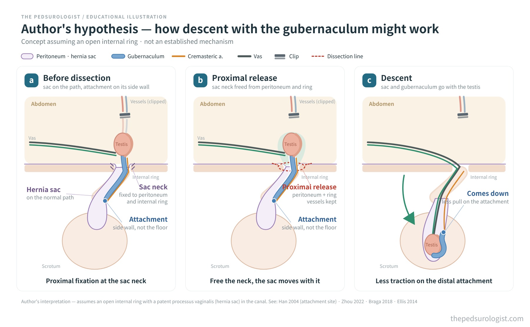
How far the evidence goes
Most data on gubernacular preservation are retrospective, and few studies have a comparison group. Because the studies compare different things with different end-points, the table is grouped by type of study. Where groups are compared, the sparing group is listed first.
| Study | Design · population | End-point · follow-up | Result |
|---|---|---|---|
| Fowler–Stephens (testicular vessels divided) | |||
| Wang 2004 | Laparoscopic two-stage, gubernaculum preserved — first description | — | Success 93% (as cited by Ellis 2014) |
| Robertson 2007 | Two-stage, laparoscopic second stage · 25 testes | Testicular survival, 6 and 18 months | Survival 88% (22/25); no comparison group |
| Mahomed 2012 | Laparoscopic-assisted two-stage · 16 patients | Volume (examination), mean 612 days | Volume similar to the contralateral side except one judged hypoplastic; no comparison group |
| Ellis 2014 | Laparoscopic-assisted two-stage · 25 patients, 28 testes | Survival and size (examination), mean 3.3 years | Survival 100%; no comparison group |
| Naycı 2017 | Single-stage, gubernaculum preserved · 7 testes | Volume and position, mean 14 months | 5 (71.4%) normal, 2 atrophic |
| Braga 2019 | Laparoscopic, allocation by surgeon preference, single- and two-stage · 212 testes | Atrophy (nubbin or impalpable, Doppler-confirmed), 3 and 12 months | Atrophy: sparing 0.6% (1/166) vs divided 28.3% (13/46) |
| Roy 2020 | Laparoscopic two-stage · 96 testes | Atrophy and re-ascent, median 12.5 months | Trend to atrophy with gubernacular division (OR 3, 95% CI 0.97–9.3, p=0.06) |
| Zhou 2022 | Open vs laparoscopic second stage, all sparing · 205 testes | Atrophy (impalpable or nubbin, ultrasound), mean 59.8 months | Atrophy 1.5%, ascent 2.9%; no non-sparing control group |
| Sacco Casamassima 2026 | Laparoscopic, single-centre retrospective · 260 testes analysed | Atrophy | Vessels preserved 6.2%, single-stage 19.3%, two-stage 10.7%; with the gubernaculum preserved, no significant difference was found between single- and two-stage |
| Randomised trials comparing sparing and division | |||
| Zhang 2021 | Randomised pilot, laparoscopic orchiopexy (high undescended testis), age 1–3 years · 120 analysed | Doppler PSV, EDV, RI and testicular volume, 1, 3, 6 and 12 months | No significant difference between groups; handling of the testicular vessels could not be confirmed from the public record |
| Abbasion 2024 | Randomised, open, within 2 cm of the internal ring · 75 of 105 testes (64 of 92 boys) analysed | Position (scrotal or high scrotal) plus volume loss <20%, 6 months; intraoperative direct measurement, ultrasound at follow-up | Anatomical success 97.2%* vs 82.1%; overall success 61.1% vs 25.6% |
| Pooled analysis | |||
| Jiryis 2026 | Meta-analysis · 3 studies, 195 patients | Atrophy | OR 0.07 (95% CI 0.01–0.91), I² 80.8%; includes orchiopexy with preserved testicular vessels, so not an estimate specific to Fowler–Stephens |
Some comparative studies reported results favouring preservation, but a randomised trial found no difference in blood flow or testicular growth. Populations, techniques and end-points differ, so the results cannot be generalised as one, and much stands in the way of comparing studies directly or reading them causally. The largest comparative study (Braga 2019) allocated by surgeon preference, with only 46 testes in the division group. Selection bias also has to be considered. A testis that can be brought down along the internal ring and canal is usually one with a patent ring, a developed gubernaculum and adequate length — a relatively less severe case. In more severe cases the ring may be closed or the gubernaculum poorly developed, so preservation itself is difficult and division with a new route is chosen. In studies where the approach is chosen on operative findings, milder testes may gather in the sparing group, and part of the difference in atrophy may reflect the testis rather than the technique (the author's view). The meta-analysis includes only three studies with heterogeneity above 80%, and definitions of atrophy vary between studies (from an impalpable testis or nubbin to a set percentage of volume loss).
This article reviews two randomised trials. The trial by Abbasion et al. studied open conventional orchiopexy without division of the testicular vessels and cannot be transferred directly to Fowler–Stephens. It reported results favouring preservation, but loss to follow-up (75 of 105 testes analysed), a baseline difference in testicular volume (significantly larger in the excision group), and the change from direct intraoperative measurement to ultrasound at follow-up have to be taken into account. All excision cases used the new route, whereas the route varied in the sparing group with the attachment site, so the effect of preservation cannot be fully separated from that of the route. A critical letter to the editor and the authors' reply were published (Aithal et al., 2024; Mottaghi et al., 2024).
By contrast, the laparoscopic randomised pilot trial by Zhang et al. (120 analysed) found no significant difference between groups in Doppler indices or testicular volume from before surgery to 12 months after (Zhang et al., 2021). It does not prove equivalence, but it is a reminder that not all results favour preservation.
The EAU guidelines (2026 edition, undescended testes section) mention gubernacular preservation in a single line. The study cited (Dave et al., 2009) did not compare preservation directly; it observed more atrophy after laparoscopic second-stage surgery when the vas formed a long loop (5 of 6 cases).
Where to use it — the author's view
In theory, preservation should make the greatest difference in operations that divide the testicular vessels — single-stage and two-stage Fowler–Stephens. Having lost its testicular artery, the testis relies on the remaining collaterals, so whether it depends mainly on the deferential system or also keeps the cremasteric / gubernacular system matters. The most natural application seems to be the second stage of a two-stage procedure, after collaterals have had time to develop. A report that, with the gubernaculum preserved, no significant difference in atrophy was found between single- and two-stage procedures (Sacco Casamassima et al., 2026) is interesting, but it is single-centre and retrospective.
I doubted whether it was needed in conventional orchiopexy, where the testicular vessels are preserved: with the main artery intact, keeping one more collateral should add little. Yet the randomised trial by Abbasion et al. reported better results with preservation in exactly that setting. Meanwhile, the laparoscopic randomised trial by Zhang et al., in which the handling of the testicular vessels is hard to confirm from the public record, found no significant difference in blood flow or testicular growth. With different populations and end-points, this question is best left open for now.
If trying it, a few things are worth checking beforehand.
- Is the internal ring patent, is the gubernaculum developed, and where is it attached? — If the ring is closed, the gubernaculum poorly developed or attached proximal to the internal ring (inside the abdomen), the route through the canal may not be feasible.
- How much peritoneum can be kept around the vas, and how much more can be freed? — The extent to which the vas and surrounding tissue can be dissected is the main limiting factor.
- The canal route is longer than the new route — there are both the view that descent through the canal raises the risk of re-ascent and reports finding no difference (discussed in Zhou et al., 2022). Position after the second stage deserves separate follow-up.
Some modifications of the traction (Shehata) technique also include gubernacular preservation (Aljneibi et al., 2026). Preserving the gubernaculum is less a technique confined to Fowler–Stephens than a principle that can be added to several ways of bringing the testis down.
Open questions
- The Fowler–Stephens studies reviewed here cannot establish an independent effect of gubernacular preservation.
- No data separate whether any benefit comes from the arteries (cremasteric), the venous return (gubernacular veins) or both.
- Definitions of atrophy and follow-up differ between studies, making direct comparison of figures difficult.
- Data comparing preservation and division with adjustment for severity — ring patency, gubernacular development, testicular position and size — are lacking.
- The extent of proximal dissection and the handling of the distal attachment are not standardised, and handling when the internal ring is closed is not described separately.
- The comparative studies reviewed here are insufficient to judge differences in long-term endocrine function or fertility.
References
- Raman JD, Goldstein M. Intraoperative characterization of arterial vasculature in spermatic cord. Urology. 2004;64(3):561-564. doi:10.1016/j.urology.2004.04.007
- Sampaio FJ, Favorito LA, Freitas MA, Damião R, Gouveia E. Arterial supply of the human fetal testis during its migration. J Urol. 1999;161(5):1603-1605. PubMed 10210426
- Benzi TC, Logsdon NT, Sampaio FJB, Favorito LA. Testicular arteries anatomy applied to Fowler-Stephens surgery in high undescended testis — a narrative review. Int Braz J Urol. 2022;48(1):8-17. doi:10.1590/S1677-5538.IBJU.2021.99.11
- Mirilas P, Mentessidou A. Microsurgical subinguinal varicocelectomy in children, adolescents, and adults: surgical anatomy and anatomically justified technique. J Androl. 2012;33(3):338-349. doi:10.2164/jandrol.111.013052
- Pascual JA, Villanueva-Meyer J, Salido E, Ehrlich RM, Mena I, Rajfer J. Recovery of testicular blood flow following ligation of testicular vessels. J Urol. 1989;142(2 Pt 2):549-552. doi:10.1016/s0022-5347(17)38812-2
- Pascual JA, Villanueva-Meyer J, Rutgers JL, et al. Long-term effects of prepubertal testicular vessel ligation on testicular function in the rat. J Urol. 1990;144(2 Pt 2):466-468. doi:10.1016/s0022-5347(17)39491-0
- Han WK, Kim JH, Hong CH, Han SW. Structural evidence against hormonal therapy for cryptorchid testis: abnormal gubernacular attachment. J Urol. 2004;171(6 Pt 1):2427-2429. doi:10.1097/01.ju.0000125271.29809.d0
- Hutson JM, Li R, Southwell BR, Newgreen D, Cousinery M. Regulation of testicular descent. Pediatr Surg Int. 2015;31(4):317-325. doi:10.1007/s00383-015-3673-4
- Wang KS, Shaul DB. Two-stage laparoscopic orchidopexy with gubernacular preservation: preliminary report of a new approach to the intra-abdominal testis. Pediatr Endosurg Innov Tech. 2004;8(3):250-253.
- Robertson SA, Munro FD, Mackinlay GA. Two-stage Fowler-Stephens orchidopexy preserving the gubernacular vessels and a purely laparoscopic second stage. J Laparoendosc Adv Surg Tech A. 2007;17(1):101-107. doi:10.1089/lap.2006.0565
- Mahomed A, Adams S, Islam S. Initial success with gubernacular-sparing laparoscopic-assisted Fowler-Stephens orchidopexy for intra-abdominal testes. J Laparoendosc Adv Surg Tech A. 2012;22(2):192-194. doi:10.1089/lap.2011.0130
- Ellis R, Lahiri R, Mahomed A. Mapping testicular blood supply in gubernaculum-sparing second-stage Fowler-Stephens procedure. Surg Endosc. 2014;28(11):3158-3161. doi:10.1007/s00464-014-3574-y
- Yamataka A, Kobayashi H, Okada Y, Tsukamoto K, Lane GJ, Miyano T. A novel technique for reinforcing the neourethra in hypospadias associated with bilateral undescended testis: use of the gubernaculum. J Pediatr Surg. 2003;38(12):1799-1801.
- Naycı A, Bahadır GB, Erdoğan C, Taşkınlar H. Laparoscopic one-stage Fowler-Stephens orchiopexy preserving gubernaculum. Turk J Urol. 2017;43(1):75-78. doi:10.5152/tud.2016.32392
- Braga LH, Ramesh S, McGrath M. Second stage gubernaculum sparing laparoscopic orchidopexy (GSLO). Societies for Pediatric Urology Fall Congress 2018, abstract. spuonline.org
- Braga LH, Farrokhyar F, McGrath M, Lorenzo AJ. Gubernaculum testis and cremasteric vessel preservation during laparoscopic orchiopexy for intra-abdominal testes: effect on testicular atrophy rates. J Urol. 2019;201(2):378-385. doi:10.1016/j.juro.2018.07.045
- Roy C, Cullis PS, Clark C, Munro FD. Retrospective analysis of testicular outcomes following laparoscopic two-stage Fowler Stephens orchidopexy. J Pediatr Surg. 2020;55(2):300-303. doi:10.1016/j.jpedsurg.2019.10.030
- Zhou G, Chen J, Yin J, et al. Open versus laparoscopic gubernaculum-sparing second-stage Fowler-Stephens orchiopexy for intra-abdominal testis: a long-term study. J Laparoendosc Adv Surg Tech A. 2022;32(8):920-924. doi:10.1089/lap.2021.0843
- Abbasion R, Mottaghi M, Saeidi Sharifabad P, et al. Gubernaculum-sparing orchiopexy versus gubernaculum excision; a randomized trial on success and atrophy rates. J Pediatr Urol. 2024;20(5):969-976. doi:10.1016/j.jpurol.2024.06.041
- Aithal S, Sinha A, Rathod KJ, Saxena R. Letter to editor: Gubernaculum-sparing orchiopexy versus gubernaculum excision. J Pediatr Urol. 2024;20(6):1229. doi:10.1016/j.jpurol.2024.08.024
- Mottaghi M, Abbasion R, Rezaei MM. Response to the "Letter to editor: Gubernaculum-sparing orchiopexy versus gubernaculum excision". J Pediatr Urol. 2024;20(6):1230-1231. doi:10.1016/j.jpurol.2024.09.005
- Mohamed A, Macdonald CM, Keene D, Marshall D, et al. Two-stage Fowler-Stephens orchidopexy in management of undescended testes: is it time for a change? A UK multi-centre retrospective study. J Pediatr Surg. 2025;60(12):162665. doi:10.1016/j.jpedsurg.2025.162665
- Sacco Casamassima MG, Saylors S, Noel-Macdonnell JR, et al. Outcomes of laparoscopic orchiopexy for intra-abdominal testis: a single institutional experience. J Pediatr Surg. 2026;61(3):162877. doi:10.1016/j.jpedsurg.2025.162877
- Zhang J, Yang X, Zhang C, et al. Testicular blood supply and growth in children with high cryptorchidism treated with gubernaculum preserving vs. cutting laparoscopic orchiopexy: a pilot trial. J Pediatr Urol. 2021;17(6):835.e1-835.e6. doi:10.1016/j.jpurol.2021.10.002
- Jiryis T, Lopes RI, Suartz CV, et al. A comparative meta-analysis and systematic review of gubernaculum sparing versus gubernaculum excision orchidopexy. J Pediatr Surg. 2026;61(8):163180. doi:10.1016/j.jpedsurg.2026.163180
- Dave S, Manaboriboon N, Braga LH, et al. Open versus laparoscopic staged Fowler-Stephens orchiopexy: impact of long loop vas. J Urol. 2009;182(5):2435-2439. doi:10.1016/j.juro.2009.07.050
- Aljneibi A, Safoury HS, Hobeldin M, Abdelsalam S, et al. Modified testicular traction in intra-abdominal testes, do we really need to wait for 6 weeks? J Pediatr Urol. 2026;106049. doi:10.1016/j.jpurol.2026.106049
- EAU Guidelines on Paediatric Urology, 2026 edition — Undescended testes. EAU Guidelines Office, Arnhem.