
After cardiac anomalies, hydronephrosis is the most common abnormality detected on prenatal ultrasound. It is found in 1–2% of all pregnancies (UTD consensus), and reported rates range from 0.6% to 5.4% depending on diagnostic criteria and gestational age. It is also a common reason for referral to pediatric urology around the time of birth. Yet hydronephrosis is a finding, not a diagnosis. The observation that the collecting system looks dilated is the same, but behind it lie quite different conditions, from transient dilation that will disappear on its own to obstruction that needs surgery to protect renal function. The word hydronephrosis alone does not tell us the cause.
So what the clinician wants from a single ultrasound comes down to two things: is this child on the path toward surgery or loss of renal function, and if not, how long do we need to watch? Over the past 30 years, several grading systems have been built to answer these questions. This article sets out what each system measures and what it misses, and how well the grade actually predicts the natural history. Which postnatal tests to perform, and in whom, is covered in Part 2.
Hydronephrosis is a finding, not a diagnosis
The distribution of causes of antenatal hydronephrosis varies widely between reports, but the overall picture is consistent. Depending on the report, more than half is transient or physiological dilation, and the most common pathological cause is ureteropelvic junction obstruction (UPJO). Mild hydronephrosis seen in the neonatal period is often explained as resolving as ureteral peristalsis matures, but not all transient dilation can be explained by this single mechanism.
Scroll horizontally to see the full figure
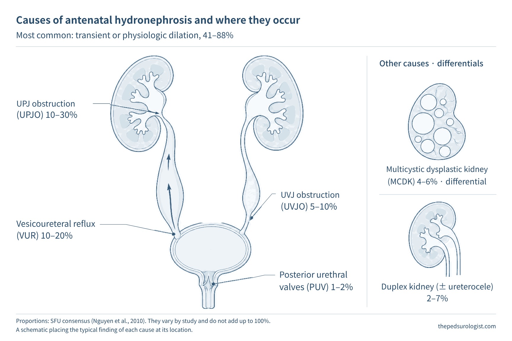
There is one point to take from this figure. Grading systems do not distinguish between these causes. The same grade contains both transient dilation and obstruction that will need surgery, and the grade only indicates the likelihood of one or the other.
What a good grading system needs
Grading systems serve to standardize description between examiners and to identify children who need further testing and follow-up. The goal is to reduce unnecessary testing and radiation exposure without missing necessary evaluation.
A good grading system should meet three conditions. First, it should predict prognosis — whether surgery will be needed, whether renal function will decline, when the dilation will resolve. Second, it should yield the same grade regardless of who measures. Third, where possible, it should use the same language before and after birth. The systems below meet these three conditions in different proportions.
Scroll horizontally to see the full figure
APD — the simplest number
The anteroposterior diameter (APD) of the renal pelvis is the front-to-back width of the pelvis measured on a transverse section of the kidney. It is measured as the maximal anteroposterior diameter of the pelvis within the renal contour on a transverse section at the renal hilum (the level where the renal vessels enter), and is distinguished from a measurement of the widest part of an extrarenal pelvis lying outside the kidney. Being a single number, it is easy to learn, objective to record, and usable both prenatally and postnatally. By UTD criteria, prenatal dilation is defined as ≥4 mm at 16–27 weeks of gestation and ≥7 mm from 28 weeks, and the threshold after 48 hours of life is ≥10 mm. However, even when the APD is below threshold, abnormalities of the calyces, parenchyma, ureter or bladder determine the risk.
Its weaknesses are also clear. The value varies with the operator and the imaging plane, and with the child's hydration status, how full the bladder is, and whether the child is prone or supine. More fundamentally, the same number means different things depending on the shape of the pelvis. An extrarenal pelvis, lying outside the kidney, may have a large APD with normal calyces and parenchyma; conversely, an intrarenal pelvis may already have markedly dilated calyces without a large APD. One of the reasons Onen proposed a new grade in 2007 was precisely the observation that APD misses severity in an intrarenal pelvis.
Yet there are Korean data showing that APD is actually better at predicting time to resolution. Jung et al. (2020) at Seoul Asan Medical Center analyzed 831 patients and 1,028 renal units with isolated hydronephrosis and showed that SFU grade and APD grade were discordant in 46.8%. Of renal units with SFU grade 2–4 but APD under 10 mm, 84.7% resolved, whereas only 53.2% of renal units with SFU grade 1 but a large APD resolved. The authors concluded that when APD is under 10 mm, SFU grade has little effect on resolution. In the study by Elmaci and Dönmez (2019) of 276 patients with isolated hydronephrosis and APD ≤20 mm, APD under 10 mm resolved at a median of 5 months and 10–20 mm at 11 months.
SFU — the most widely used grade
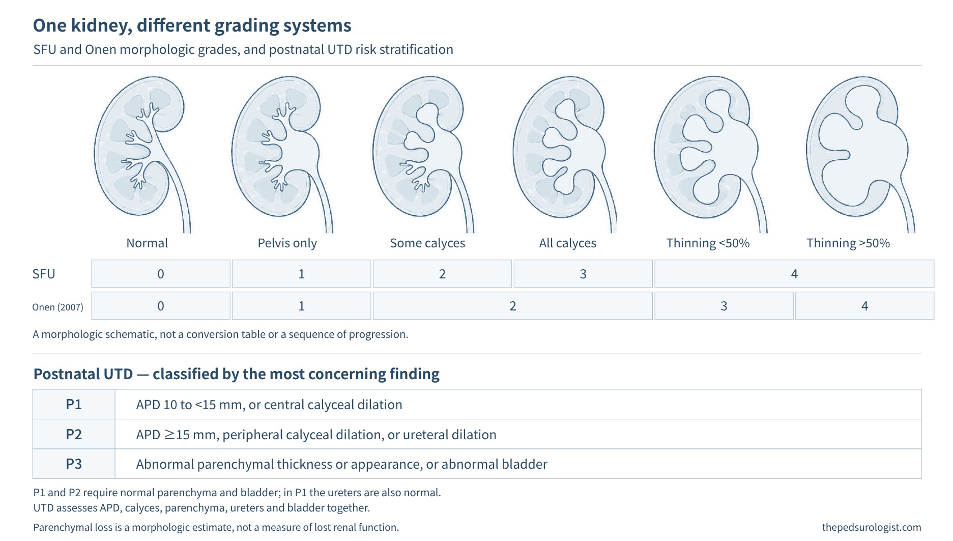
The SFU grade, published by the Society for Fetal Urology in 1993, looks at shape rather than a number. Grade 0 is no dilation, grade 1 is when only the renal pelvis is visualized, grade 2 is dilation of the pelvis and some calyces, grade 3 is dilation of all calyces, and grade 4 adds parenchymal thinning. It remains the most widely used, and most natural history data are reported in this grade.
However, there is considerable room for subjectivity in assignment. Grades 1 and 2 are separated by whether some calyces appear dilated, but the criteria are vague as to how much separation of the renal sinus counts as normal and from what point subtle calyceal dilation should be read as dilation. The bigger problem is grade 4. A kidney with mild medullary thinning and a kidney with paper-thin parenchyma fall into the same grade 4, so the kidneys that actually need surgery are hard to pick out within the grade. It is therefore important to describe the individual findings of the pelvis, calyces and parenchyma alongside the grade.
Studies of interobserver agreement also expose this limitation. When Kim et al. (2013) from the Department of Radiology at Severance Children's Hospital had four radiologists grade 180 kidneys twice each, overall interobserver agreement for SFU was substantial (κ 0.61–0.68), but agreement was low for grades 1 and 2.
Onen — splitting parenchymal loss
Onen's alternative grade (2007) kept the SFU framework but redrew the boundaries. Grade 1 is dilation of the pelvis only, grade 2 adds calyceal dilation, grade 3 is parenchymal loss of less than half, and grade 4 is loss of more than half (a cyst-like appearance with almost no visible renal parenchyma). Instead of separating calyceal dilation, it groups it into one grade and splits the parenchymal loss that SFU groups into a single grade 4 into two. Here, half refers to morphologically assessed parenchymal loss, not a corresponding reduction in function. In the original study, which followed 162 neonates and 228 renal units, kidneys at SFU grade 2 or below on the first postnatal ultrasound all resolved without functional decline, while 68.6% of SFU grade 4 kidneys underwent surgery. Differential renal function of grade 4 kidneys was scattered from 7% to 39%. The higher grades needed to be subdivided more finely. Other variants that split SFU grade 4 into 4A and 4B or use a 1–5 scale go in the same direction.
Severance Hospital has used the Onen grade since the 2010s, and two sets of data underpin this. In the Kim et al. (2013) study above, interobserver agreement for Onen (κ 0.66–0.76) was slightly higher than for SFU, but the difference was not statistically significant, and agreement was low for Onen grades 2 and 3. Lee et al. (2013) from the Department of Pediatric Urology compared renal biopsy findings obtained at pyeloplasty with preoperative indices in bilateral UPJO, where differential renal function alone makes it difficult to assess the degree of injury on each side. In 29 renal units, the degree of histological injury (glomerulosclerosis, interstitial fibrosis, dysplastic change) was not associated with differential renal function or SFU grade, but was clearly associated with the Onen grade. In bilateral disease, differential renal function is the relative proportion between the two kidneys and can mask deterioration on both sides together; in that setting, subdividing parenchymal loss reflected histological injury. However, this is a result from a small surgical cohort, and it cannot be extended to evidence that Onen is better for all hydronephrosis.
That said, Onen did not change the nature of grading. It is still the appearance on a single ultrasound, it does not distinguish causes, and it does not look at the ureter and bladder. This is why, even in a center that records Onen grades, ureteral and bladder findings must be documented separately.
UTD — a consensus bridging prenatal and postnatal
In 2014, eight US societies (four in imaging, plus pediatric urology, fetal urology, pediatric nephrology and maternal–fetal medicine) came together to agree on a new classification. This is the urinary tract dilation (UTD) classification. It looks at six parameters — anteroposterior renal pelvic diameter, calyceal dilation (central and peripheral), parenchymal thickness, parenchymal appearance, ureter, and bladder. Prenatally, unexplained oligohydramnios is added. And it classifies prenatal and postnatal findings within the same framework. There is one principle of assignment: the most concerning finding among the findings determines the risk group. Even if APD is in the P1 range, peripheral calyceal dilation makes it P2; if there is urinary tract dilation with a parenchymal or bladder abnormality, it is P3 even if APD is under 15 mm. Transient visualization of the ureter postnatally is not by itself classified as ureteral dilation.
Scroll horizontally to see the full figure
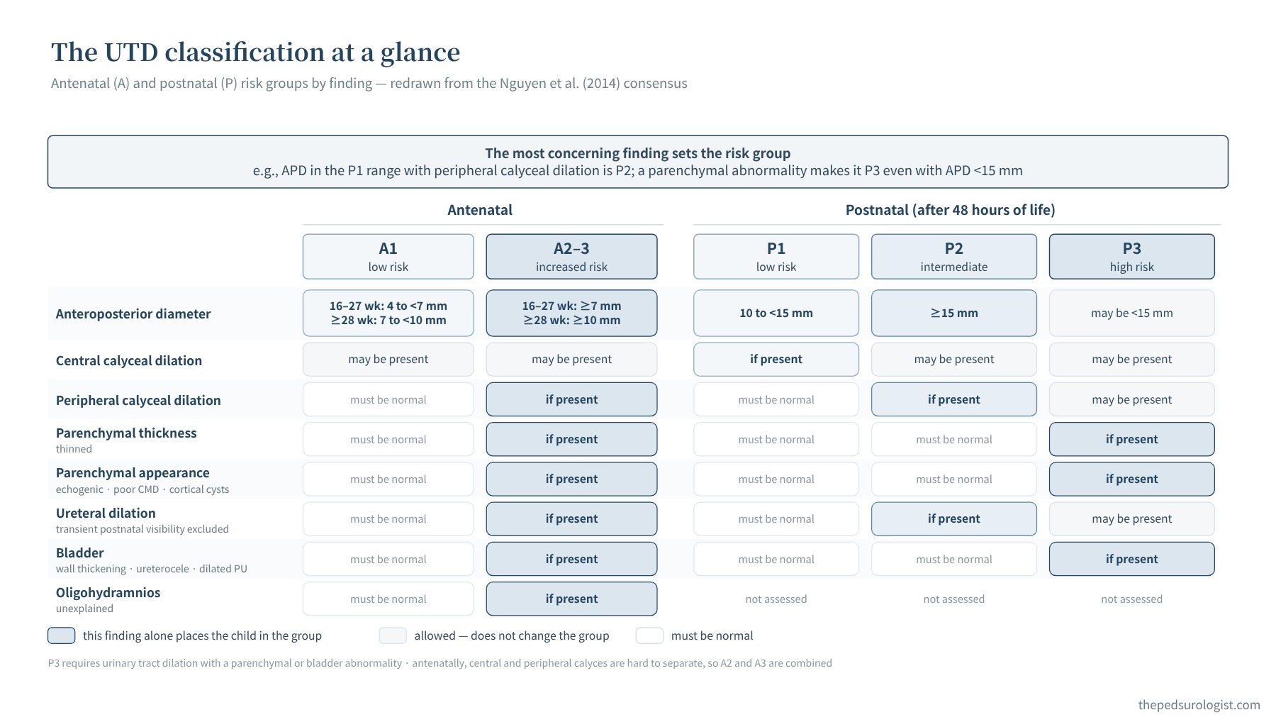
UTD has three strengths. The language used prenatally can be carried over unchanged after birth, the ureter and bladder are included in the grade, and each grade comes with a follow-up plan. Hodhod et al. (2016) in Montreal regraded 730 renal units by SFU and UTD and reported that UTD is reliable (parallel-forms reliability 0.92) and that UTD risk group independently predicts surgery, and SFU grade independently predicts resolution.
But the remaining problems are also clear. First, causes are mixed within the same risk group. Second, the distinction between central and peripheral calyces is prone to vary between examiners, and the consensus itself notes that the two are difficult to distinguish prenatally, particularly before the third trimester. Third, there is no numerical criterion for judging parenchymal thinning. Fourth, renal growth is not taken into account. In a study by a Brazilian group that followed 447 patients with isolated hydronephrosis for a median of 9 years (Melo et al., 2022), all systems predicted the need for surgery well (AUC 0.90 or higher), but prediction of kidney injury was more accurate with a modified classification adding parenchymal thickness measurements to UTD (AUC 0.913 vs 0.887). This suggests that quantifying the parenchyma can add to prediction; further validation in other cohorts is needed before the modified classification can be used in routine practice.
How well does the grade predict the natural history?
There are many data on natural history, but most cover isolated hydronephrosis as a whole, including transient dilation and not only UPJO, and the population, whether surgical patients are included, and the definition of resolution differ between studies. So even the overall resolution rate ranges widely, from 64–75% in meta-analyses to 25–55% in recent observational studies (AAP 2025 summary). The direction is consistent. Mild dilation mostly resolves on its own, and the higher the grade, the slower and less often it resolves. The same resolution rate cannot be attached to every grade.
Four representative studies show this difference. Sidhu et al. (2006) systematically reviewed 25 studies of isolated antenatal hydronephrosis and meta-analyzed 7 of them, grouping grades into 1–2 and 3–4 and reporting the proportion "stable or improved" rather than resolved (98%, 51%). Braga et al. (2018), in a prospective cohort of 401 patients out of 855 after excluding surgery, late referral, no postnatal dilation and associated anomalies, defined resolution as APD ≤10 mm; 3-year cumulative resolution was 98% for SFU grade 1, 87% for grade 2, 76% for grade 3 and 57% for grade 4 (by UTD, P1 90%, P2 81%, P3 71%). Elmaci and Dönmez (2019), in 276 patients with isolated hydronephrosis and APD ≤20 mm, excluding SFU grade 4 on the first postnatal ultrasound, defined resolution as SFU 0 with APD under 7 mm. Over a median follow-up of 16 months, 71.7% resolved, the median time to resolution was 6 months, and there were no operations. Jung et al. (2020), in 1,028 renal units with isolated hydronephrosis at a single Korean center, defined resolution as APD under 5 mm with SFU grade 1, and 48-month resolution rates were 81.7% for grade 1, 65.6% for grade 2, 37.6% for grade 3 and 5.2% for grade 4.
Scroll horizontally to see the full figure
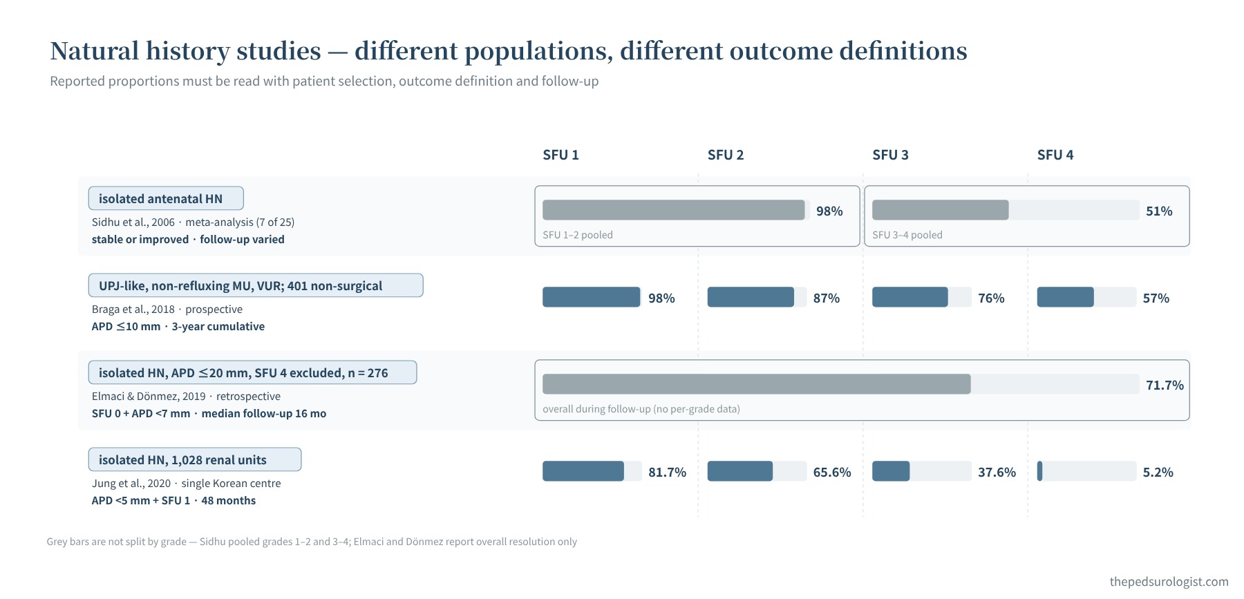
Before setting the numbers in the figure side by side, one must first check what was counted as resolution, who was analyzed, and at what time point the number applies. The same SFU grade 4 is 57% in the Braga study and 5.2% in the Jung study. The former is a 3-year cumulative resolution rate in a cohort that excluded children who had surgery, with resolution defined as APD ≤10 mm; the latter is a 48-month resolution rate using a far stricter definition, APD under 5 mm with SFU grade 1. Because population selection, definition and time point all differ, the gap cannot be attributed to any single one of them. When quoting numbers to parents, it is important to keep in mind the conditions behind them.
The conclusion of Braga et al. (2018) is practical. In both systems, higher grades resolved more slowly and predictive performance was similar, so the authors considered that whichever system is used, counseling parents about the timing of resolution can be the same. However, a number from one system cannot be transferred to the same-numbered grade of another. In the meta-analysis by Lee et al. (2006), which predicted postnatal pathology from the prenatal grade, the risk of a pathological cause confirmed after birth rose to 11.9%, 45.1% and 88.3% for mild, moderate and severe prenatal dilation, respectively. In contrast, the risk of vesicoureteral reflux did not differ clearly by degree of dilation. This is why selecting children for VCUG, which Part 2 covers, cannot be based on the grade alone.
The course over time, not a single grade
In summary, current grading systems are fairly good at identifying who will go on to surgery, but are less accurate about when resolution will occur and whose kidneys will be silently injured. And no system tells us the cause. The initial grade is a starting point for estimating risk, and what revises that judgment is change during follow-up. Whether the dilation increases, whether the parenchyma thins, whether the kidney grows appropriately, and how the ureter and bladder look are reassessed repeatedly under the same conditions, and functional studies and symptoms are read alongside when needed.
Hydronephrosis that improved and then causes problems again years later, and when to end follow-up, are covered in a separate article.
The hydronephrosis grade indicates likelihood, not cause. SFU, Onen and UTD carry different information, so numbers are not transferred one-to-one between them, and natural history figures are quoted only after checking the population and the outcome definition. What revises the judgment is not a single grade but change during follow-up.
Next — When should the first postnatal ultrasound be done? Who should have a voiding cystourethrogram (VCUG) and diuretic renography, and who should receive continuous antibiotic prophylaxis? We read the UTD consensus (2014), the American Academy of Pediatrics clinical report (2025), the EAU/ESPU guidelines (2026), and the data behind them.
References
- Kim SW. Management of hydronephrosis: a comprehensive review in pediatric urology perspective. Child Kidney Dis. 2024;28(2):59-65. doi:10.3339/ckd.24.011
- Nguyen HT, Herndon CDA, Cooper C, et al. The Society for Fetal Urology consensus statement on the evaluation and management of antenatal hydronephrosis. J Pediatr Urol. 2010;6(3):212-231. doi:10.1016/j.jpurol.2010.02.205
- Sinha A, Bagga A, Krishna A, et al. Revised guidelines on management of antenatal hydronephrosis. Indian Pediatr. 2013;50(2):215-231. doi:10.1007/s13312-013-0064-6
- Lee RS, Cendron M, Kinnamon DD, Nguyen HT. Antenatal hydronephrosis as a predictor of postnatal outcome: a meta-analysis. Pediatrics. 2006;118(2):586-593. doi:10.1542/peds.2006-0120
- Nguyen HT, Benson CB, Bromley B, et al. Multidisciplinary consensus on the classification of prenatal and postnatal urinary tract dilation (UTD classification system). J Pediatr Urol. 2014;10(6):982-998. doi:10.1016/j.jpurol.2014.10.002
- Fernbach SK, Maizels M, Conway JJ. Ultrasound grading of hydronephrosis: introduction to the system used by the Society for Fetal Urology. Pediatr Radiol. 1993;23(6):478-480. doi:10.1007/BF02012459
- Onen A. An alternative grading system to refine the criteria for severity of hydronephrosis and optimal treatment guidelines in neonates with primary UPJ-type hydronephrosis. J Pediatr Urol. 2007;3(3):200-205. doi:10.1016/j.jpurol.2006.08.002
- Kim SY, Kim MJ, Yoon CS, Lee MS, Han KH, Lee MJ. Comparison of the reliability of two hydronephrosis grading systems: the Society for Foetal Urology grading system vs. the Onen grading system. Clin Radiol. 2013;68(9):e484-e490. doi:10.1016/j.crad.2013.03.023
- Lee YS, Jeong HJ, Im YJ, et al. Factors indicating renal injury in pediatric bilateral ureteropelvic-junction obstruction. Urology. 2013;81(4):873-878. doi:10.1016/j.urology.2012.09.064
- Jung J, Lee JH, Kim KS, Park YS. Utility of Society for Fetal Urology and anteroposterior pelvic diameter grading systems for estimating time to resolution of isolated hydronephrosis: a single center study. J Urol. 2020;204(5):1048-1053. doi:10.1097/JU.0000000000001140
- Elmaci AM, Dönmez Mİ. Time to resolution of isolated antenatal hydronephrosis with anteroposterior diameter ≤ 20 mm. Eur J Pediatr. 2019;178(6):823-828. doi:10.1007/s00431-019-03359-y
- Hodhod A, Capolicchio JP, Jednak R, et al. Evaluation of urinary tract dilation classification system for grading postnatal hydronephrosis. J Urol. 2016;195(3):725-730. doi:10.1016/j.juro.2015.10.089
- Melo FF, Vasconcelos MA, Mak RH, et al. Postnatal urinary tract dilatation classification: improvement of the accuracy in predicting kidney injury. Pediatr Nephrol. 2022;37(3):613-623. doi:10.1007/s00467-021-05254-x
- Sidhu G, Beyene J, Rosenblum ND. Outcome of isolated antenatal hydronephrosis: a systematic review and meta-analysis. Pediatr Nephrol. 2006;21(2):218-224. doi:10.1007/s00467-005-2100-9
- Braga LH, McGrath M, Farrokhyar F, Jegatheeswaran K, Lorenzo AJ. Society for Fetal Urology classification vs urinary tract dilation grading system for prognostication in prenatal hydronephrosis: a time to resolution analysis. J Urol. 2018;199(6):1615-1621. doi:10.1016/j.juro.2017.11.077
- Herndon CDA, Otero HJ, Hains D, et al. Perinatal urinary tract dilation: recommendations on pre-/postnatal imaging, prophylactic antibiotics, and follow-up: clinical report. Pediatrics. 2025;156(1):e2025071814. doi:10.1542/peds.2025-071814
- EAU Guidelines on Paediatric Urology. Dilatation of the upper urinary tract (PUJ obstruction); Foetal urology. European Association of Urology, 2026.