Before measuring the angle in a hypospadias repair, there is a prior question. What is holding the penis down? Two boys with similar ventral curvature may differ entirely: in one it straightens once the skin and subcutaneous tissue are released, in the other curvature persists after a thorough dissection. The visible angle is the same; the structure that needs correcting is not.
Before answering that question, it is worth examining the words we use. The two terms used most often in this field — chordee and corporal disproportion — each suggest a mechanism. The names help us think and plan, but they do not identify the causative tissue in a given patient.
What the names tell us, and what they do not
Chordee comes from Latin chorda and, behind that, Greek χορδή — a cord, a string, the gut-string of a lyre. It entered English through the French cordée, meaning corded.
The word did not enter medicine to describe a congenital anomaly. Older dictionaries define chordee as a painful, downward-curving erection accompanying gonorrhoea. The idea of the time — that inflamed, indurated ventral tissue pulls the erection down — is preserved inside the word. As the term came to be applied to congenital curvature across the first half of the twentieth century, the mechanism it suggests travelled with it.
That said, the point should not be overstated. Chordee today does not necessarily presuppose a real cord. Devine himself noted in the 1990s that chordee denotes curvature and does not designate the causative tissue. The same applies to corporal disproportion, discussed below. The etymology and the convention evoke a particular mechanism, but the clinical name alone does not establish which tissue is responsible in a given patient. The names are useful for understanding curvature and planning an operation; they do not name the culprit.
What is holding it down — separating the contributing components
Four components can be separated out, and several usually act together in the same patient. They are not, however, four tissues stacked in order. The dartos is superficial fascia and Buck's fascia is deep fascia, so grouping them under one heading does not mean they lie at the same depth. And because degloving releases the skin and the superficial fascia together, the surgical sequence alone cannot fully separate what each tissue contributes.
Scroll horizontally to see the full figure
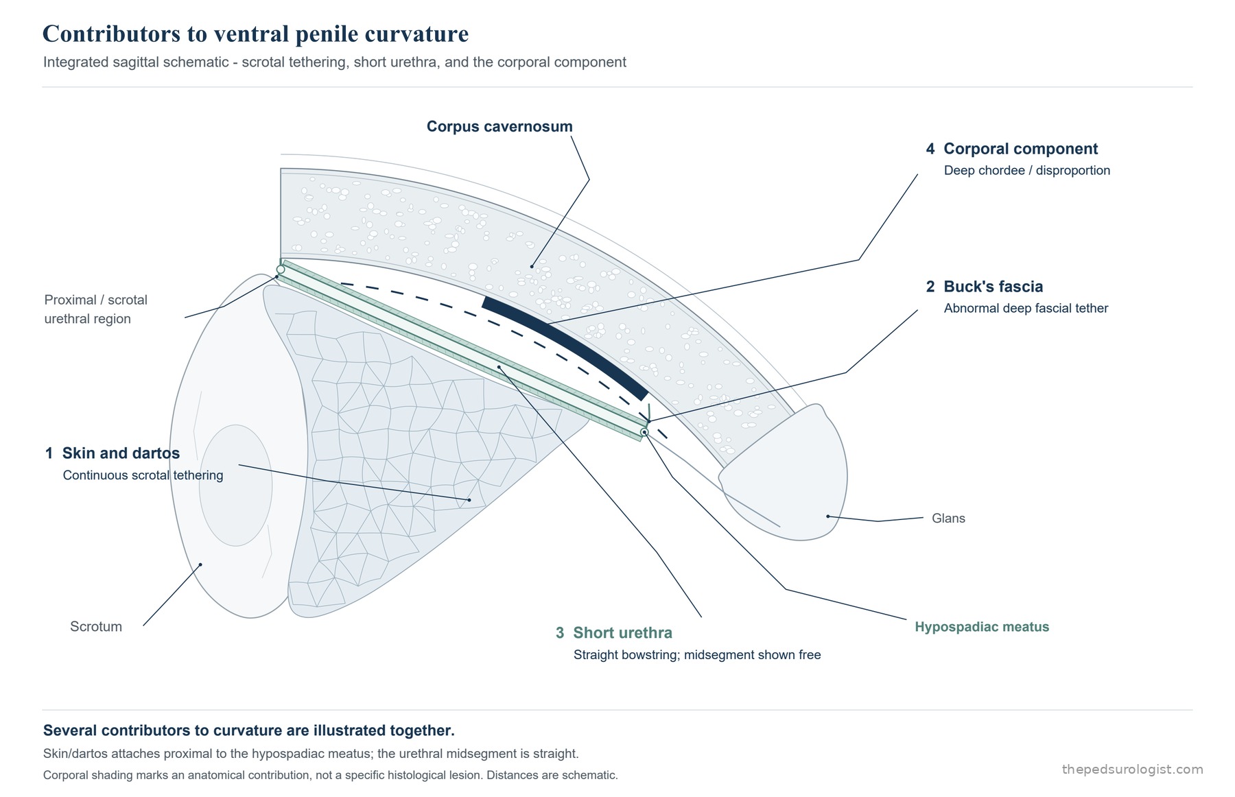
| Component | What contributes to the curvature | What to check intraoperatively |
|---|---|---|
| 1 Skin | Tethering from insufficient ventral skin length or abnormal fixation and adhesion | How much straightening follows degloving |
| 2 Dartos and Buck's fascia | Tethering by inextensible, dysplastic fascial tissue | Whether the curvature improves after dissection |
| 3 Urethra / urethral plate | Tethering or hinging from short length or insufficient extensibility | Whether curvature from the urethra remains after the surrounding tissue is dissected |
| 4 Corporal factor | Restraint from an abnormality of the tunica itself (deep chordee), or an imbalance in relative dorsal–ventral length and extensibility | Whether curvature persists after the external tethering and urethral factors have been assessed and released |
Most superficially, the ventral skin is short or abnormally fixed and holds the penis down. This tethering runs up continuously from the scrotum and attaches proximal to the meatus. These are the cases in which degloving alone substantially reduces the curvature. A penis that looked markedly bent at the start may need little deeper correction once the skin is released. The angle measured before the skin is taken down is therefore a poor basis for choosing a correction. Degloving is both exposure and a way of gauging the cause — though the skin and the superficial fascia are released together at this step, so the straightening gained cannot be credited to the skin alone.
Next comes fascial tethering, where dysplastic dartos and Buck's fascia form inextensible ventral tissue that holds the penis down. The two fasciae lie at different depths — the dartos superficial, Buck's deep — but in ventral dissection they often come away as a single block, and for separating causes it is clearer to group them. This tissue converges toward the splayed corpus spongiosum and attaches densely to the ventral tunica albuginea. It is not so much that the splayed spongiosum pulls, as that this is where the dysplastic fascia meets the tunica. The hypoplastic spongiosum is part of the ventral defect and is excised in severe cases, but it is hard to regard it as the agent of tethering.
The ventral dartos also differs histologically from normal — though the two studies do not report the same thing. In 100 patients whose excised ventral dartos was examined, collagen thickness and degree of fibrosis correlated significantly with the severity of curvature (p=0.002 and 0.017). In a comparison of 50 boys with hypospadias against 50 age-matched controls, collagen density was instead reduced. On the amount of collagen the two reports diverge.
What they point to together is not quantity but composition. In the latter study elastin (ELN) expression was reduced and the COL3A1:COL1A1 ratio was lower — less of the distensible type III collagen, relatively more of the stiffer type I. Both changes point to a matrix that does not stretch. So tissue that has lost its elasticity fits these data better than tissue thickened like scar, and the degree of that change tracks the severity of the curvature.
This is correlation, not proof of causation. But set alongside the operative fact that excising this tissue straightens the penis, it is reason to regard the ventral dartos as genuinely taking part in the tethering. The abnormality is not uniform either — the proximal dartos was the most consistently abnormal, while in distal hypospadias with mild curvature the distal dartos was indistinguishable from controls. The dartos is both a lesion to be removed and the material that will cover the neourethra, so how much to excise depends on location and on the degree of curvature.
Third, the urethra and urethral plate may limit ventral length through short length or insufficient extensibility. The two are not the same thing. The urethra is the tube proximal to the meatus; the urethral plate is the strip of epithelium that never became a tube, running along the ventral surface from the meatus to the glans. It is the plate that is incised and rolled into a tube in TIP, while the rarely reported "urethral tethering" in Donnahoo's classification (7%) refers to a congenitally short or hypoplastic urethra. They may also restrain the ventral side in different ways, so which of them is responsible has to be checked separately.
In severe hypospadias in particular the plate can act as a short hinge, preventing the corpora from extending at erection. The presence of a plate is not in itself a cause, however. The plate rarely produces curvature in distal hypospadias. What matters is not the appearance of the plate but whether it still restricts ventral length after the deep tissue has been dissected. This distinction bears directly on the plan: the plate is tissue available for urethral reconstruction, but if it is also the structure maintaining the curvature, then preservation and correction come into conflict.
The fourth factor is given its own section below.
Where the quoted proportions come from. The four-category classification in common use derives from studies of congenital curvature without hypospadias. Among the 87 patients of Donnahoo and colleagues, skin tethering accounted for 32%, fibrotic dartos and Buck's fasciae 33%, corporeal disproportion 28% and urethral tethering 7%; in the 102 cases of Polat and colleagues the figures were roughly 39, 30, 27 and 4%. The main categories are similar but the proportions are not identical, and above all these are not hypospadias cohorts. That premise should be kept in mind when applying them.
How the explanations and the surgical strategies widened
| Period · source | What is documented |
|---|---|
| 18th c. → early 20th c. | Chordee, used for the painful curvature of gonorrhoea, comes to be applied to congenital curvature |
| 1973 Devine & Horton | Distinguish developmental anomalies of the periurethral structures and fasciae; release the dysplastic fascial and connective tissue while preserving the urethra where possible |
| 1975 Kaplan & Lamm | Ventral curvature observed in 89% of 46 fetal specimens; propose arrest of development |
| 1982 Kramer et al. | Corporeal disproportion the largest group, 9 of 20; the same paper concludes that most cases arise from anomalies of the tissue layers |
| 1980s Duckett et al., flap era | The urethral plate itself is regarded as dysplastic tissue causing the curvature, so it is divided and the urethra rebuilt with a preputial flap (transverse preputial island tube) — the standard for proximal cases |
| 1994 Baskin & Duckett | Among 374 boys undergoing onlay island flap repair, 50 (13%) still had significant curvature after degloving, mostly mild (20–30°) corporal disproportion — the shift back to preserving the plate: onlay replaces tube (10%→33%), residual curvature corrected by dorsal plication |
| mid-1990s onward Snodgrass, TIP era | TIP spreads and plate preservation becomes standard; the anatomical and histological studies of 1998–2000 add to its rationale |
| 2009 Snodgrass & Prieto | Extend preservation through urethral and plate mobilisation, with additional correction for residual curvature |
| 2016–2022 Tissue-characterisation studies | Camoglio reports increased stiffness of the corpus spongiosum; Hadidi describes excision of the outer longitudinal layer of the tunica in correcting deep chordee |
| 2017–2021 Snodgrass & Bush | Re-evaluate the limits of plate preservation in proximal hypospadias — the plate's own tethering is the problem after all — and emphasise staged reconstruction |
This table should not be read as one explanation replacing another. The periods overlap because the debate genuinely ran in parallel. Kramer's single 1982 paper contains both corporeal disproportion and anomalies of the tissue layers, and the discussions of the mid-1990s set out primary growth imbalance, secondary corporal asymmetry from external deforming forces, and dysplastic tissue side by side. No explanation disappeared and then returned.
It is worth noting that the view of the urethral plate has come full circle. In the 1980s the plate itself was taken to be dysplastic tissue that produced the curvature, so it was divided. In the 1990s, the observation that it often was not the cause moved practice toward preserving it. Since the 2010s the view has come back round: in severe proximal cases the plate does turn out to be holding the ventral side down. The place it returned to is not the place it started, though — the question now is not whether plates tether, but which plate does.
One thing to add. What the 1998–2000 studies biopsied was the urethral plate and the tissue immediately beneath it. That no fibrous band was found there does not mean there is no abnormal tissue anywhere on the ventral side. Deeper down, the outer longitudinal layer of the tunica is tissue those studies never looked at.
The observation of Kaplan and Lamm is also worth holding on to. That 89% of fetal penises were ventrally curved means curvature is normal in fetal life and resolves on its own before birth. If so, the curvature in hypospadias may be not something that appeared but a straightening that did not run to completion. What interrupts that process, this study did not establish.
The name corporal disproportion
When curvature persists on the erection test after the external tethering has been fully released, we call it corporal disproportion — meaning the ventral aspect is short relative to the dorsal.
This name has its limits too. In practice disproportion is usually not a measurement but a diagnosis of exclusion: the urethra has even been divided, curvature remains, therefore it is a length difference. Within the literature I was able to search, studies that actually measured dorsal and ventral tunical length and demonstrated a difference were difficult to find.
Operative findings suggest something else may occupy that position. Hadidi reports that correcting curvature in perineal hypospadias required excising not only the hypoplastic urethral plate and the atretic corpus spongiosum but the outer longitudinal layer of the tunica albuginea, and gives this entity the name deep chordee, using it as the branch point for choosing a technique. This is not external tethering tissue but a layer of the covering of the corpora.
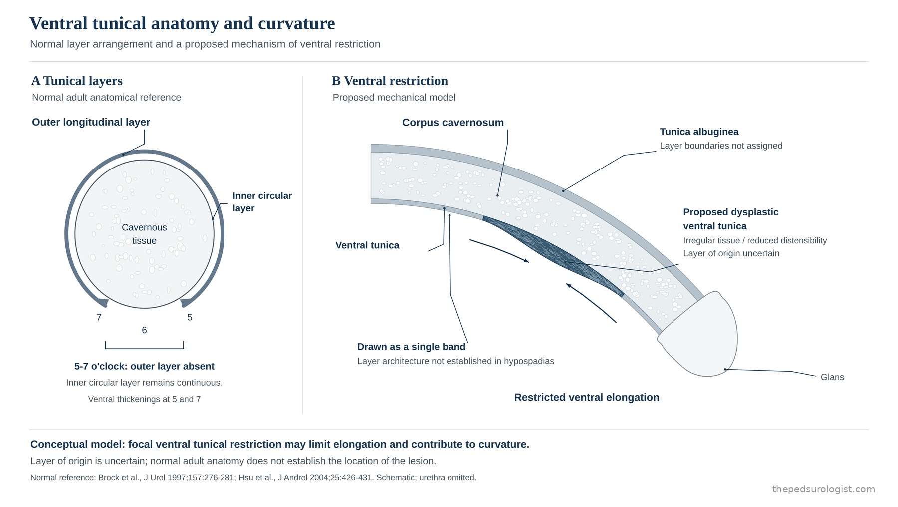
For anatomical background, the tunica albuginea of the normal adult corpus cavernosum is bilayered, and the fibres of the outer longitudinal layer are absent over the ventral midline. The inner circular layer is maintained even there, and at the 5- and 7-o'clock positions sit triangular structures continuous with the anterior fibres of the bulbospongiosus (the ventral thickening). The ventral midline is where the tunica is most atypical even in normal anatomy.
This is, however, normal adult anatomy. Hypospadias is precisely a failure of that ventral side to form, so the absence of the outer layer in normal men cannot be transferred to conclude that there is no such layer to excise — nor can its presence be asserted. Normal anatomy does not establish where the lesion lies. Hadidi's excision of the outer layer should be taken as a separate surgical-anatomical observation.
Scroll horizontally to see the full figure
Histological reports of the ventral tissue do exist. Ekmark and colleagues analysed urethral plate and deep chordee biopsies from 14 boys with hypospadias and found the deep chordee to be connective tissue containing coarse nerve bundles, smooth muscle bundles and vessels of irregular wall thickness. That the endothelium was WT-1 negative suggests this tissue is malformed from the outset rather than scar or a reparative response. In tissue excised from 17 boys with severe curvature, Sennert, Hadidi and colleagues likewise found sparse elastic fibres, disorganised smooth muscle and abnormal blood sinusoids.
And yet the word tunica does not appear in these reports. Ekmark's description never uses the term tunica albuginea, and does not state whether the biopsy came from the point of maximal curvature. That does not make the tunica irrelevant — at the same site, Hadidi describes removing the hypoplastic plate, the atretic corpus spongiosum and a layer of the tunica as a single block.
Set the three descriptions side by side and a common feature appears. The tissue here does not divide cleanly along normal layer lines. Connective tissue, smooth muscle, coarse nerves and malformed vessels are intermingled; elastic fibres are sparse and the smooth muscle is disorganised; and at operation the plate, the spongiosum and the tunica come away together. On that reading, it is closer to the findings to regard this as a single dysplastic tissue in which hypoplastic corpus spongiosum and tunica are blended than to assign three separate structural names to it.
This is an interpretation, not a reported finding: tunical tissue has not been histologically identified in any of those specimens. But if the interpretation holds, the question changes shape — from "which layer is diseased" to "why did this region fail to differentiate into layers at all." Given that hypospadias is a failure of ventral development, it is expecting the normal layer arrangement at the ventral midline that sits awkwardly. Here, once again, a name has settled the mechanism in advance.
In short, I could not find data that labels the ventral tunica at the point of maximal curvature and answers directly what it is made of or which layer it comes from. What the reports above analysed was the urethral plate and the deep tissue beneath it; no study appears to have characterised the excised tunical layer separately, and the fetal specimen reported as having a normal tunica was a single distal hypospadias. This cannot be stated as an absence from the whole literature — but data answering the question directly remains hard to find.
That the corporal factor is real, and is the thing to be corrected, is shown by surgical outcomes. Snodgrass and Bush report 43 boys with proximal hypospadias and curvature of 30° or more after degloving (mean 70°) in whom the urethral plate was transected and, in the 65% with curvature still remaining, ventral corporotomy was performed — with no recurrence. There is plainly a share that persists once the external tethering and the urethra have both been dealt with, and that share has to be addressed at the corpora.
What has to be distinguished is how each of these contributes: inextensible ventral tissue, tethering by the urethra and urethral plate, an abnormality of the tunica itself, and the relative dorsal–ventral length relationship. Observations on physiological fetal curvature and subsequent straightening offer a developmental perspective, and the operative findings in deep chordee show the deep tissue that is actually the target of correction. A mechanistic account linking the two is possible, but the evidence is not sufficient to explain all curvature by a single tissue or a single developmental process.
The starting point for understanding penile curvature in hypospadias is not a single angle but the structures that produce it. Skin, fascia, the urethra and urethral plate, the corpora — four contributors in differing measure, several of them at once in the same boy. The names we use help us separate them, but they do not point to the tissue responsible. Chordee presupposes a cord, corporal disproportion a difference in length, deep chordee a particular layer — all three carry inside the name a mechanism that has never been confirmed. What is actually met deep on the ventral side is a single dysplastic tissue that does not divide cleanly into layers, and what that tissue is has yet to be established histologically. Judgement during the operation therefore has to rest on stepwise observation rather than on names — checking, at each stage, how much straightening followed what was released is for now the surest way to separate the causes. The next question — when correction is needed, and how much residual curvature is acceptable — belongs to the next article.
Next in this series — Hypospadias, Part 7 — How Much Curvature to Correct, and in What Order: where the 30° threshold is actually used, artificial erection and measurement, and the stepwise path from plate preservation to transection.
References
- Devine CJ Jr, Horton CE. Chordee without hypospadias. J Urol. 1973;110(2):264-271. doi:10.1016/s0022-5347(17)60183-6
- Kaplan GW, Lamm DL. Embryogenesis of chordee. J Urol. 1975;114(5):769-772. doi:10.1016/s0022-5347(17)67140-4
- Kramer SA, Aydin G, Kelalis PP. Chordee without hypospadias in children. J Urol. 1982;128(3):559-561. doi:10.1016/s0022-5347(17)53045-1
- Baskin LS, Duckett JW, Ueoka K, Seibold J, Snyder HM. Changing concepts of hypospadias curvature lead to more onlay island flap procedures. J Urol. 1994;151(1):191-196. doi:10.1016/s0022-5347(17)34915-7
- Baskin LS, Duckett JW. Dorsal tunica albuginea plication for hypospadias curvature. J Urol. 1994;151(6):1668-1671. doi:10.1016/s0022-5347(17)35341-7
- Baskin LS, Erol A, Li YW, Cunha GR. Anatomical studies of hypospadias. J Urol. 1998;160(3 Pt 2):1108-1115. doi:10.1097/00005392-199809020-00039
- Donnahoo KK, Cain MP, Pope JC, et al. Etiology, management and surgical complications of congenital chordee without hypospadias. J Urol. 1998;160(3 Pt 2):1120-1122. doi:10.1097/00005392-199809020-00041
- Baskin LS, Erol A, Li YW, Liu WH. Anatomy of the neurovascular bundle: is safe mobilization possible? J Urol. 2000;164(3 Pt 2):977-980. doi:10.1097/00005392-200009020-00014
- Snodgrass W, Patterson K, Plaire JC, Grady R, Mitchell ME. Histology of the urethral plate: implications for hypospadias repair. J Urol. 2000;164(3 Pt 2):988-989. doi:10.1097/00005392-200009020-00017
- Hsu GL, Hsieh CH, Wen HS, et al. Anatomy of the human penis: the relationship of the architecture between skeletal and smooth muscles. J Androl. 2004;25(3):426-431. doi:10.1002/j.1939-4640.2004.tb02810.x
- Tang Y, Huang J, Chen S, Huang L, Wang M. Anatomical features of congenital chordee without hypospadias in children and implication for diagnosis and treatment. Zhongguo Xiu Fu Chong Jian Wai Ke Za Zhi. 2006;20(3):217-219.
- Polat EC, Erdem MR, Topaktas R, Ersoz C, Onol SY. Our experience in chordee without hypospadias: results of 102 cases. Urol J. 2014;11(4):1783-1787.
- Camoglio FS, Bruno C, Zambaldo S, Zampieri N. Hypospadias anatomy: elastosonographic evaluation of the normal and hypospadic penis. J Pediatr Urol. 2016;12(4):199.e1-199.e5. doi:10.1016/j.jpurol.2016.02.007
- Snodgrass W, Bush N. Staged tubularized autograft repair for primary proximal hypospadias with 30-degree or greater ventral curvature. J Urol. 2017;198(3):680-686. doi:10.1016/j.juro.2017.04.019
- Hadidi AT. Perineal hypospadias: back to the future Chordee Excision & Distal Urethroplasty. J Pediatr Urol. 2018;14(5):424.e1-424.e9. doi:10.1016/j.jpurol.2018.08.014
- Nozohoor Ekmark A, Grelaud D, Hansson E, Svensson H, Arnbjörnsson E, Gisselsson D. The cellular architectures of hypospadias. Pediatr Dev Pathol. 2020;23(6):476-478. doi:10.1177/1093526620943084
- Sennert M, Perske C, Wirmer J, Fawzy M, Hadidi AT. The urethral plate and the underlying tissue; a histological and histochemical study. J Pediatr Urol. 2022;18(3):364.e1-364.e9. doi:10.1016/j.jpurol.2022.02.006
- Duarsa GWK, Tirtayasa PMW, Winarti NW, Michael A, Duarsa KHA. Correlation between histopathology properties of dartos tissue and the severity of penile curvature in hypospadias. Arch Ital Urol Androl. 2024;96(4):12894. doi:10.4081/aiua.2024.12894
- Raharja PAR, Birowo P, Rachmadi L, et al. Comparative analysis of collagen density and COL1A1, COL3A1, and ELN expression in dartos fascia of hypospadias patients. J Pediatr Urol. 2025;21(6):1572-1580. doi:10.1016/j.jpurol.2025.06.021
- Snodgrass W, Prieto J. Straightening ventral curvature while preserving the urethral plate in proximal hypospadias repair. J Urol. 2009;182(4 Suppl):1720-1725. The account of changing surgical strategy also draws on Campbell-Walsh-Wein chapter 56 and the ESPU guideline. The etymology follows the chordee entries in the Century Dictionary and GCIDE. Literature searching was performed in PubMed.