We usually explain reflux starting at the bladder. Urine travels the wrong way, and infection reaches the kidney with it. None of that is wrong. But there is a patient the explanation does not reach.
The newborn whose kidney is already small, who has never had a urinary tract infection. Picked up on antenatal hydronephrosis, with cortical defects on a DMSA scan taken in the first days of life. No infection, no outlet obstruction.
That kidney was not damaged by the reflux. It was made alongside it, in the same event. And that event happens in the fourth week of gestation.
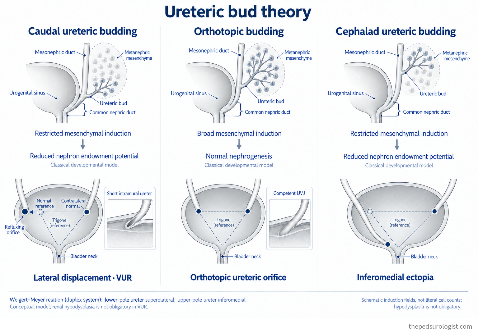
So this note follows reflux from the mesonephric duct rather than from the bladder. Where the ureteric bud arises sets the position of the orifice, the length of the submucosal tunnel, and the quality of the kidney — all at once.
First, primary and secondary
One distinction has to be made before anything else, because the same word covers two different things.
- Primary — a congenital defect of the ureterovesical junction itself: reflux occurs despite an adequately low-pressure storage profile in the bladder. Campbell adds a line worth keeping — it is not simply a lack of tunnel length. A dilated ureter, a ureteral wall that does not collapse readily, weak detrusor backing and the characteristics of the bladder all play a part.
- Secondary — something else overwhelms the junction, classically sustained or repeated rises in intravesical pressure.
| Group | What it covers |
|---|---|
| Primary | A congenital defect of the junction itself — short intramural tunnel, a dilated ureter, a ureteral wall that resists collapse, weak detrusor backing |
| Other congenital anomalies of the junction | Ureteral duplication, ureterocele, paraureteral diverticulum — classified differently by different sources (see below) |
| Secondary — pressure | From the bladder (neurogenic, non-neurogenic) · from the outlet (mechanical obstruction such as posterior urethral valves, stricture or a mass, and functional obstruction) |
| Iatrogenic | Surgery or injury involving the junction |
Campbell's phrase intrinsic stiffness of a ureter means a ureteral wall that does not collapse readily. The finite-element work below points the same way — a stiffer or thicker ureteral wall required more pressure to collapse. But that effect reached significance only at wall thicknesses not seen in practice (a 2.5 mm wall in a 6.4 mm ureter). Plausible as a mechanism, then, but not something the model demonstrated across the clinical range. The dilated ureter is a separate item: it is the one that prompts long tunnels or ureteral tapering and plication at reimplantation.
Where to put ureteral duplication, ureterocele and the paraureteral diverticulum depends on the source. The current Campbell chapter groups them under secondary reflux, as an "anatomically abnormal UVJ". The long-standing classification, and several other textbooks, place them on the primary side, usually as a separate heading — primary reflux associated with other congenital anomalies of the junction.
Both have a case. Reading them as secondary stresses that a junction that could have worked was undermined by an adjacent lesion; reading them as primary stresses that the junction itself is congenitally abnormal. Since the mechanism is plainly different from pressure-driven secondary reflux, keeping them in a group of their own — as in the table above — causes least confusion in practice.
This note is about primary reflux. Embryology explains the primary form; in secondary reflux the same junction is overwhelmed by pressure or by an adjacent lesion, and those are the protagonists. The two do not separate cleanly in clinic, though. Campbell says as much — primary and secondary reflux "may not always be mutually exclusive," and what is perceived as primary may in fact carry a secondary component, as with bladder overactivity or bladder–bowel dysfunction. Which is why treatment begins with normalising bladder and bowel function: strip out the secondary element and what remains is the true primary disease.
A kidney requires a meeting
Around the fourth week the ureteric bud sprouts from the caudal mesonephric duct and grows into the metanephric blastema. It branches dichotomously as it extends cranially, and the ampulla at each branch tip induces the surrounding mesenchyme to condense and convert to epithelium. That epithelium becomes the nephron.
Each generation of branches becomes collecting duct; the first five or so are remodelled and dilated into the calyces and renal pelvis. One side builds the plumbing and the other the filtering units — and neither can build its half alone.
That is the premise of everything below. A kidney exists only where the bud and the blastema meet. Mice lacking Pax2 or Wt1 form no ureteric bud at all, and their mesenchyme fails to respond to induction even when recombined with a normal bud. Without Wnt4 the bud invades but tubulogenesis stops there.
Which raises the question the rest of this note turns on. What happens when the bud meets the wrong part of the blastema?
How the orifice finds its place
At five to six weeks the segment of mesonephric duct left below the origin of the bud — the common excretory duct, also called the common nephric duct — dilates and connects to the urogenital sinus. This is not a simple confluence: it proceeds through apoptosis, which detaches the ureter from the mesonephric duct so that it can fuse with the bladder separately. Vitamin A signalling is thought to mediate the step.
As incorporation proceeds the ureteric orifice migrates cranially and laterally across the bladder base, while the mesonephric duct orifice travels caudally and medially to become the ejaculatory duct. One Y-shaped stem is absorbed and its two arms are drawn in opposite directions.
The picture of a stem being "absorbed into the bladder" is best taken as a classical model that aids understanding. What actually happens involves apoptosis and tissue rearrangement — and the origin of the trigone, below, departs from the classical picture too.
The fibromuscular wall of the trigone was long taught to form by extension of the ureter into the bladder (Weiss, 1988). Lineage tracing in mice by Viana and colleagues in 2007 found that trigonal muscle arises mostly from bladder smooth muscle, with only a minor contribution from mesenchyme associated with the ureter — Campbell sets the two side by side and calls it a contrast to the classic hypothesis. Any diagram that explains the trigone as "ureter that moved inside the bladder" needs revising.
When the bud arises off-target
One thing should be said plainly first. The sequence above has not been observed directly in humans. It is inferred, primarily from the natural experiment of duplex kidneys.
In a duplex system the upper pole orifice rotates dorsally relative to the lower pole orifice and comes to lie more caudally and medially. Weigert and Meyer recognised the regularity of that relationship, and it carries their names as the Weigert–Meyer rule. For the rule to hold, there must be a fixed relationship between where the bud arises and when it is absorbed — and applying that relationship to a single system gives the following.
- A bud arising closer to the urogenital sinus than normal (caudally) → the common excretory duct left below it is short, so it is absorbed early → over-rotation draws the orifice cranially and laterally → intramural and submucosal length fall short → reflux.
- A bud arising more cranially → a long common excretory duct, incompletely absorbed → insufficient rotation leaves the orifice caudal and medial → it opens at or below the bladder neck → ectopia and obstruction.
So far this is a story about the ureter. The decisive step Mackie and Stephens added in 1975 was this: the same misplacement also changes the contact between bud and metanephric blastema. A bud arising outside the normal zone fails to meet the appropriate part of the blastema, and the consequence is dysplasia and hypoplasia — in the extreme, agenesis.
Hypoplasia is too little development — a reduced nephron number. Dysplasia is abnormal differentiation and organisation of the tissue itself. They often travel together but they are not synonyms, and imaging that shows a "small kidney" does not by itself say which one it is.
So one misplacement produces two things at once — a refluxing orifice and a kidney that was never fully built. They are siblings, not cause and effect.
Swipe sideways to read the whole diagram
Why the tunnel matters — and is 5:1 a law?
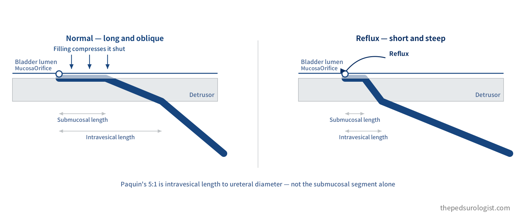
The intramural ureter is compressed passively as the bladder fills. There is no separate active valve: given enough intramural length and fixation, rising intravesical pressure closes the ureter against itself. During voiding, longitudinal muscle within the tunnel contracts and draws the extravesical and intravesical points together, shortening and widening it so the bolus can pass. The same structure opens and closes according to the moment.
Swipe sideways to read the whole diagram
The numbers come from Paquin's dissections of the ureterovesical junction in children in 1959.
| Age | Intravesical ureteral length | Submucosal length | Ureteral diameter at the junction |
|---|---|---|---|
| 1–3 yr | 7 mm | 3 mm | 1.4 mm |
| 3–6 yr | 7 mm | 3 mm | 1.7 mm |
| 6–9 yr | 9 mm | 4 mm | 2.0 mm |
| 9–12 yr | 12 mm | 6 mm | 1.9 mm |
In non-refluxing junctions the ratio of tunnel length to ureteral diameter was roughly 5:1; in refluxing junctions, 1.4:1. That contrast has been the design brief of every reimplantation for sixty years.
The tunnel length in question is the intravesical length, not the submucosal segment alone. Dividing 7 mm by 1.4 mm at 1–3 years gives exactly five; dividing the submucosal 3 mm gives a little over two. Conflate the two and the same "5:1" describes an entirely different operation.
The table says one more thing. Submucosal length grows from 3 mm to 6 mm while ureteral diameter barely moves. The ratio improves on its own with growth. Spontaneous resolution is attributed to just this — remodelling of the junction over time, with progressive elongation and consolidation of the intravesical ureter. It is worth noting that the decision to wait has a structural basis.
But 5:1 is not a law. As early as 1969 Lyon and colleagues argued that the configuration of the orifice matters as much as tunnel length, and recent finite-element models have come down on their side.
- Beyond a tunnel length of 1 cm, further length does not meaningfully reduce the pressure required to collapse the ureter.
- Changing the length-to-diameter ratio from 3:1 to 5:1 altered the required closing pressure by less than 7%.
- Making the orifice a 2 mm protruding "volcano" reduced it by 78%.
- And classical mechanics predicts, contrary to Paquin's intuition, that a wider ureter collapses more easily — which is what the model showed.
Antireflux competence was never going to come down to a single ratio. The shape of the orifice, the properties of the ureteral wall itself, muscular support and fixation, ureteral peristalsis, and bladder pressure and function all contribute. 5:1 is one of those factors pinned to a number, not the boundary between a normal and a refluxing junction.
None of which should be read as "Paquin was wrong." Sixty years have confirmed that operating to 5:1 gives good results. What may be wrong is our explanation of why it works. Knowing that something works is not the same as knowing the mechanism.
A different route to the same failure — the paraureteral diverticulum
So far everything has been explained by where the bud arose. But there is another route to reflux, one that does not require a short tunnel: the collapse of the other condition named in the previous section — the backing.
Scroll sideways to see the full figure
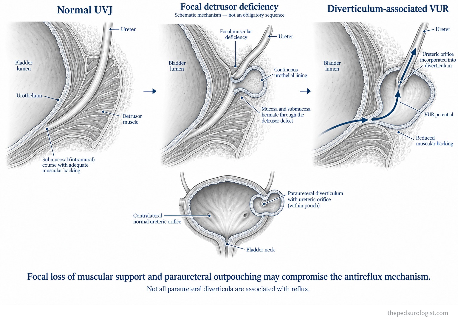
The mechanism. Where the detrusor beside the ureteric hiatus is focally deficient, mucosa and submucosa herniate through the defect into a pouch. The urothelial lining remains continuous, so from inside the bladder it looks as though a second opening has appeared beside the ureteric orifice. This is the paraureteral diverticulum, the one Hutch described.
The trouble comes as the pouch enlarges. Once the ureteric orifice is drawn into the diverticulum, the intramural ureter loses the muscle behind it — the very force that compressed it shut during filling and voiding. The tunnel is as long as it was, but there is no longer a floor to compress it against, and the length-to-diameter arithmetic of the previous section gives no answer here.
Not every paraureteral diverticulum is associated with reflux. Where the orifice remains outside the pouch and keeps its backing, many go without reflux. What matters is not the presence of the diverticulum but whether the orifice has been incorporated into it.
Congenital and acquired are different stories. A diverticulum arising from a congenital detrusor defect belongs with the "other congenital anomalies of the junction" in the table in section 1. A diverticulum produced over time by sustained high intravesical pressure — posterior urethral valves, a neurogenic bladder — is something else: the cause is pressure, so it is secondary in the sense of section 1, and the diverticulum is a consequence. The same radiographic finding can tell two different stories. It is worth noting that Hutch first described this diverticulum, in 1952, in paraplegic patients — an acquired pouch produced by high pressure. What now carries the same name in children is usually the congenital defect, so a shared eponym does not imply a shared origin.
Why it is counted separately in prognosis. A deficient backing is not the kind of problem that growth fills in. That is why the VUR Index, which we come to in Part 2, counts complete ureteral duplication and the paraureteral diverticulum together as "ureteral anomalies" — reflux accompanied by these took significantly longer to improve or resolve.
Why the kidney suffers — two routes
Here we return to the patient this note opened with. And the answer is not one thing.
Congenital dysplasia — settled before any infection
DMSA shows cortical defects in infants with high-grade reflux and no history of urinary tract infection and no outlet obstruction. These infants are mostly male, and the refluxing units cluster at grades IV and V. Data from prenatally diagnosed male infants show the distribution.
| Grade of reflux | Normal | Slight damage | Severe damage |
|---|---|---|---|
| I–III | 13 (100%) | — | — |
| IV | 8 (53%) | 5 (34%) | 2 (13%) |
| V | 2 (15%) | 5 (38%) | 6 (46%) |
The table supports the Mackie–Stephens hypothesis and at the same time draws its boundary. Grades I–III were all normal. Congenital damage is not a property of reflux in general but a property of high-grade reflux. "If there is reflux, the kidney is underbuilt" does not hold.
Nor does the converse. High-grade reflux does not mean every kidney is hypoplastic or necessarily short of nephrons — 15% of the grade V kidneys in that table were normal. Bud theory is a framework for understanding the congenital renal anomalies that accompany reflux, not a tool for estimating an individual patient's nephron number from a reflux grade.
Acquired scarring — the route that needs infection
The evidence on the other side is older and firmer. Ransley and Risdon created reflux in pigs by unroofing the intramural ureter. Pyelonephritis and reflux nephropathy followed only where the urine was subsequently infected. Without infection, no nephropathy was observed.
Hence the flat statement in the current textbook — nephropathy from primary reflux requires colonisation of the urine with pathogenic bacteria, in the face of normal voiding. Sterile reflux rarely causes meaningful upper tract deterioration on its own.
That should not be read as "infection leaves only a transient dip in function." Once pyelonephritis occurs, inflammation and tissue damage leave a permanent scar. Saying infection is necessary is a very different claim from saying its consequences are mild.
There is also a caveat. In Hodson's experiments, adding bladder outlet obstruction to reflux produced atrophic pyelonephritis without infection. Under high pressure, sterile reflux is not harmless — which is the reason treatment starts with normalising bladder and bowel function.
Timing matters too. The risk of post-infectious scarring is greatest in the first year of life and falls with age. Ransley and Risdon proposed a "big bang" theory for it — most scarring occurs at the first episode of pyelonephritis, and further scarring is unlikely without repeated episodes. Four uncertainties remain attached to it: the assumption that younger kidneys are more susceptible is more weakly substantiated than it looks; new scars were reported in the landmark reflux studies; imaging cannot distinguish post-infectious defects from intrinsic developmental dysplasia; and the appearance of a defect changes as the kidney grows.
Between 0 and 6 months the male-to-female ratio of reflux runs as high as 3:1; by 21 to 24 months it is 1:1. The first group is high-grade reflux found through antenatal hydronephrosis — the congenital dysplasia route. The second is reflux found after a urinary tract infection — the acquired scarring route. Two largely different populations sit under one diagnosis.
How far genetics has got
Reflux recurs in siblings at a high rate and clusters in families, which invites a genetic explanation. The current report card is modest.
At the budding stage RET (GDNF signalling), PAX2, EYA1 and SIX1 come up; at the metanephric stage, ROBO2, SLIT2 and SRGAP1. Overexpressing Ret in mice places the ureteric bud abnormally and raises the incidence of reflux at birth to around 30%, against 4% in wild type — a molecular footing for the very "position" Mackie and Stephens were describing. In syndromic CAKUT the gene-to-phenotype links are clear.
For non-syndromic primary reflux, they are not. Large parent–child trio analyses and case–control studies have produced no compelling association with any single gene or locus, despite autosomal dominant patterns in many families. The reading at present is that the mechanism is more complex and polygenic.
What I take from this
- Reflux is not a disease of the bladder but of how the ureterovesical junction was built. Where the bud left the mesonephric duct sets both the position of the orifice and the length of the tunnel.
- A short tunnel lengthens with growth. Submucosal length increases while ureteral diameter barely changes. The decision to wait rests on anatomy.
- 5:1 is a useful operative standard, not a proven threshold. Evidence is accumulating that orifice configuration may matter more than tunnel length.
- A small kidney in a boy with high-grade reflux may not be the result of infection. Conversely, in most reflux the kidney needs an infection in order to be harmed.
- Correcting reflux does not restore nephrons that were never formed, or scars already present. Surgery prevents future damage, not past damage.
And in practice the two routes coexist. Cortical defects on imaging often cannot be sorted cleanly into congenital maldevelopment and post-infectious scarring, which is why assessment takes in not only the grade but kidney size and structure, function, infection history and bladder function.
That last point is where conversations go wrong most often. It is why "will correcting the reflux protect the kidney?" has no single answer. Preventable damage and damage already settled sit under the same diagnosis, and unless they are separated, discussions about the goal of treatment talk past each other — with parents, and in the arguments about antibiotic prophylaxis.
Mackie–Stephens is a widely accepted model, but it is a hypothesis. This note treats it as one, and keeps what it predicts separate from what has been observed.
References
- Hutch JA. Vesico-ureteral reflux in the paraplegic: cause and correction. J Urol. 1952;68(2):457-469. doi:10.1016/S0022-5347(17)68223-5
- Khoury AE, Wehbi E. Management Strategies for Vesicoureteral Reflux. In: Campbell-Walsh-Wein Urology, 13th ed. Elsevier.
- Baskin LS, et al. Embryology of the Human Genitourinary Tract. In: Campbell-Walsh-Wein Urology, 13th ed. Elsevier.
- Mackie GC, Stephens FD. Duplex kidneys: a correlation of renal dysplasia with position of the ureteral orifice. J Urol. 1975;114(2):274-280.
- Paquin AJ. Ureterovesical anastomosis: the description and evaluation of a technique. J Urol. 1959;82:573-583.
- Lyon RP, Marshall S, Tanagho EA. The ureteral orifice: its configuration and competency. J Urol. 1969;102(4):504-509.
- Villanueva CA, Nelson CA, Stolle C. Intravesical tunnel length to ureteral diameter ratio insufficiently explains ureterovesical junction competence: a parametric simulation study. J Pediatr Urol. 2015;11(3):144.e1-5.
- Villanueva CA, Tong J, Nelson C, Gu L. Ureteral tunnel length versus ureteral orifice configuration in the determination of ureterovesical junction competence: a computer simulation model. J Pediatr Urol. 2018;14(3):258.e1-6.
- Ransley PG, Risdon RA. Reflux nephropathy: effects of antimicrobial therapy on the evolution of the early pyelonephritic scar. Kidney Int. 1981;20(6):733-742.
- Marra G, Barbieri G, Dell'Agnola CA, et al. Congenital renal damage associated with primary vesicoureteral reflux detected prenatally in male infants. J Pediatr. 1994;124(5 Pt 1):726-730.
- Batourina E, Tsai S, Lambert S, et al. Apoptosis induced by vitamin A signaling is crucial for connecting the ureters to the bladder. Nat Genet. 2005;37(10):1082-1089.
- Viana R, Batourina E, Huang H, et al. The development of the bladder trigone, the center of the anti-reflux mechanism. Development. 2007;134(20):3763-3769.
- Figures for the sex distribution of reflux and for the genetics of non-syndromic reflux follow the 2017 reports cited in chapter 41 of Campbell-Walsh-Wein Urology.