
We tell families that if a child with reflux develops a urinary tract infection, the kidney may be damaged. The sentence is familiar, but several steps have been left out of it. How do bacteria in the bladder reach the renal parenchyma, and what happens in the infected tissue? And why, after the infection has been treated with antibiotics, does a scar remain in some children and not others?
Part 1 separated the congenital renal abnormalities that accompany reflux from the acquired damage that follows infection; Part 2 looked at the likelihood of resolution and the risk carried while waiting. This article follows what lies between them — the path from infection reaching the kidney to tissue injury and scar.
Between a bladder infection and an infection of the renal parenchyma
The diagnostic label "urinary tract infection" does not by itself explain renal damage. An infection confined to the bladder and a pyelonephritis that has invaded the renal parenchyma differ in where the injury sits. To understand acquired renal scarring, the first step is the stage at which bacteria reach the kidney and provoke inflammation.
VUR offers bladder organisms a hydrodynamic route to the upper tract. Bacterial virulence — the capacity to adhere to epithelium, to multiply, and to withstand host defences — and the patient's own defences then act together. This is why infection and its course differ between children with the same grade of reflux.
But the presence of reflux, the occurrence of renal infection, and the formation of a permanent scar are not the same event. Children without reflux develop pyelonephritis and acquired scars; conversely, some children with persistent reflux grow stable kidneys and never become infected. This is the point at which Part 1 divided congenital dysplasia from acquired scarring, and this article looks inside the acquired side.
When interpreting sterile reflux, bladder pressure and function must be considered alongside it. In posterior urethral valves or a high-pressure bladder, declining renal function may be observed in the absence of infection. Congenital abnormalities of renal development and the effects of pressure and obstruction must be assessed together; the loss of function should not be read wholesale as acquired damage caused by reflux.
Equally, the effects of high pressure cannot all be attributed to congenital hypoplasia. In Hodson's postnatal porcine experiments, focal interstitial fibrosis and tubular injury developed with high pressure and intrarenal reflux even without infection. This is the basis for distinguishing normal-pressure primary VUR from situations with high pressure or obstruction.
Papillary shape changes intrarenal reflux
The idea that pressure transmitted through reflux acts directly on renal tissue was historically framed as the water hammer effect. That is a hypothesis about the direct consequence of pressure, and it must be distinguished from intrarenal reflux (IRR), the route by which infected urine enters the kidney itself.
If VUR is urine travelling from bladder to ureter to pelvis and calyx, intrarenal reflux is urine in the calyx passing backwards into the collecting ducts of the papilla. At this second boundary, the shape of the renal papilla becomes decisive.
Ransley and Risdon, examining porcine renal papillae, showed that intrarenal reflux differed according to papillary shape. In a convex, simple papilla, collecting ducts open onto the papillary surface as oblique, slit-like orifices. As calyceal pressure rises, these openings tend to close, resisting retrograde flow. In a flat or concave, compound papilla, the ducts open more nearly at right angles and more widely, so that under the same pressure the orifices do not coapt and urine passes readily into the ducts.
This is the same group as the other experiment seen in Part 1 — where reflux alone produced no nephropathy unless the urine was infected. One line of work showed that infection is necessary; the other showed where the breach occurs when infection is present.
Scroll sideways to view
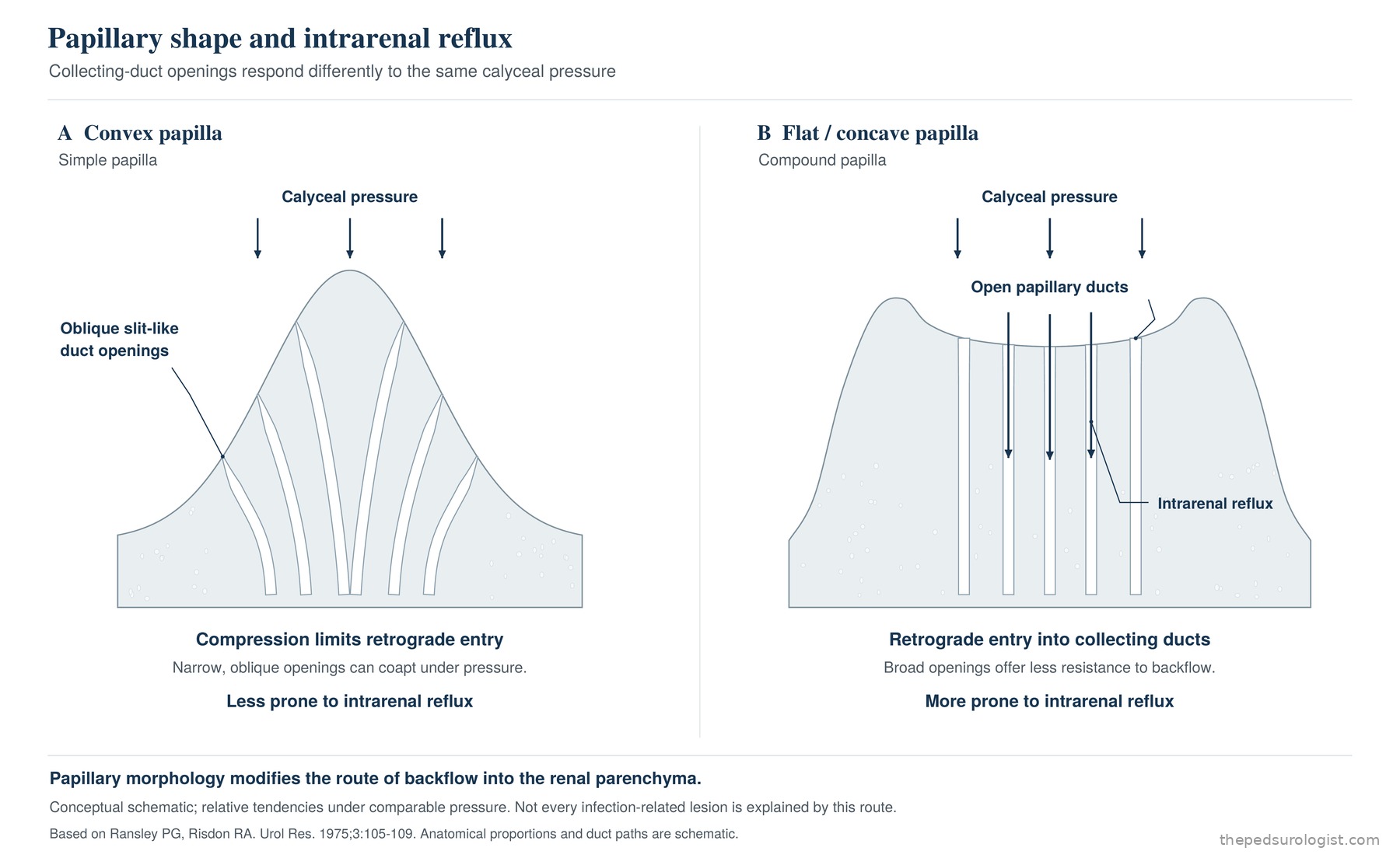
Compound papillae are relatively more frequent at the upper and lower ends of the kidney — the upper and lower poles. That intrarenal reflux and scarring are commonly seen there can be explained by this anatomy. Even with infected urine throughout the collecting system, not every region is exposed — or damaged — equally.
The route has also been documented on imaging in children. In a series of 50 children (59 renal units) in whom intrarenal reflux was seen on voiding cystourethrography, which I reported with colleagues, the sites of intrarenal reflux corresponded to the sites of photon defect on DMSA in 76.3%, and 65.2% of those defects went on to scar. Intrarenal reflux marks where contrast entered, and that territory overlaps substantially with the territory that is subsequently damaged. This was a retrospective series of children who all had intrarenal reflux, so it describes what happened in the territory where intrarenal reflux was seen rather than a risk compared with children without it.
Intrarenal reflux is an important route for infected urine to enter the renal parenchyma, but it does not account for all infection-related renal damage. The extent of actual injury also involves bacterial characteristics, the host response and microcirculation, and any accompanying functional abnormality.
The response that clears bacteria and the response that injures tissue
Renal epithelium senses pathogens and organises the surrounding defence. TLR4 recognises the lipopolysaccharide of Gram-negative organisms, and cytokines and chemokines released by the epithelium direct the migration of inflammatory cells. IL-8 and its receptors CXCR1 and CXCR2, the complement system, neutrophils and macrophages all participate.
The first purpose of this response is bacterial clearance. Organisms must be phagocytosed, their multiplication restrained and the spread of infection halted before tissue can recover. The abundance of inflammatory cells is therefore not by itself grounds for reading the whole response as harmful.
The problem arises when infection and inflammation are insufficiently controlled, or when the response fails to terminate properly in injured tissue. Bacterial epithelial injury compounded by inflammatory oedema, capillary congestion and impaired regional perfusion, oxidative stress and inflammatory mediators can amplify tubular and interstitial damage. In severely affected areas, microabscesses and tissue destruction appear, and the subsequent repair may tilt towards fibrosis rather than restoration of normal architecture.
Why the balance matters is shown well by neutrophil-depletion experiments. Ruiz-Rosado and colleagues framed this as a neutrophil–macrophage imbalance. Neutrophils were central to preventing bacterial ascent, and when they were experimentally depleted, monocytes increased compensatorily and macrophage-driven inflammation became excessive. Scarring worsened and serum creatinine, BUN and potassium rose. Clearing the bacteria and ending the inflammation on time are both necessary.
This mechanism does not imply that general anti-inflammatory treatment works. In the paediatric randomised trial by Rius-Gordillo and colleagues, adding dexamethasone did not reduce scarring — 22% versus 21% (p=0.907) among the 91 children who completed follow-up. The authors themselves note, however, that the target sample size and the expected scarring rate were not reached, so the result is read more accurately as "there is currently no basis to recommend it" than as "steroids do not work".
Practice therefore centres on controlling infection early with appropriate antimicrobial treatment, and on correcting the conditions that can perpetuate injury, such as obstruction and bladder dysfunction.
Why a scar remains after the infection is over
That the bacteria have been cleared and that the injured tissue has recovered its original architecture are two different facts. Reversible inflammation and functional decline may recover, but where tissue destruction was severe, tubules are lost, collagen accumulates in the interstitium and normal structure may be replaced by fibrous tissue. A scar is what that process of injury and repair leaves behind.
Scroll sideways to view
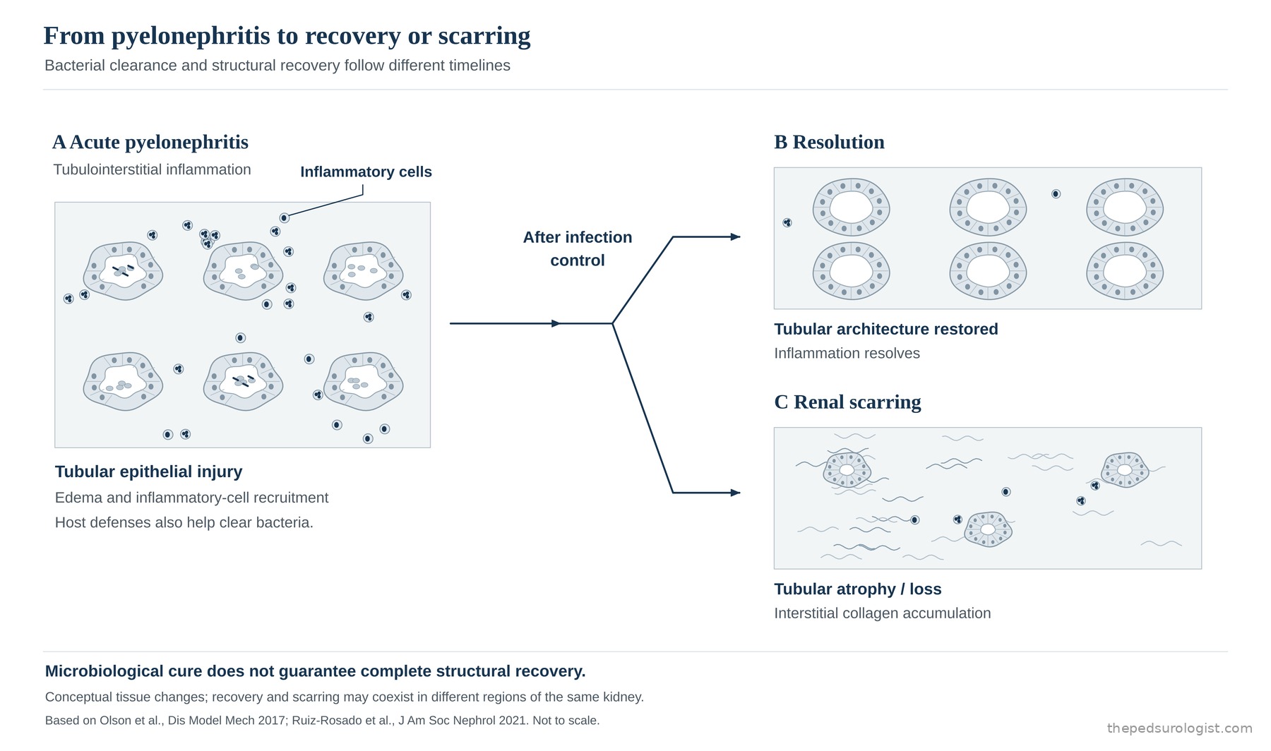
Olson and colleagues' murine work shows this directly. Five days of ceftriaxone resolved bladder and kidney infection, yet abscesses remained histologically in some animals at the end of treatment, and a month later a similar proportion showed scars with fibrosis and inflammatory infiltrate. Tissue remodelling and scar maturation can continue after microbiological cure. It follows that finding a scar on later imaging does not establish that infection persisted until then, or that antibiotic treatment failed.
One further detail in the same study is worth noting: among treated animals, renal function was preserved in almost all. That a scar remains and that function has declined do not automatically travel together. It is a murine study nonetheless, and does not transfer straightforwardly to the human course.
Scars matter functionally because the amount and architecture of working renal tissue is reduced. Outcome depends on the extent of injury and whether it is bilateral, on the function of the contralateral kidney, and on any accompanying congenital abnormality. A focal scar does not lower total renal function in every child, but extensive parenchymal loss can be linked over the long term to hypertension, proteinuria and hyperfiltration.
Also to be weighed is acquired injury added to a kidney that congenitally had less functioning tissue. A scar of the same size may carry a different meaning depending on the functional reserve the child started with.
When to assess what the kidney is left with
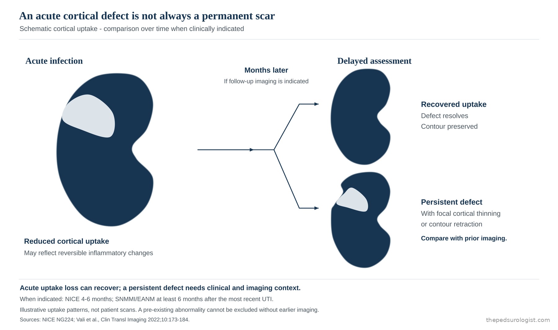
In acute pyelonephritis, DMSA uptake may be reduced by oedema, inflammation and tubular dysfunction. These defects can recover over several months, so the extent of defect in the acute phase should not be read directly as the extent of permanent tissue loss.
Scroll sideways to view
| Finding | What to weigh in interpreting it |
|---|---|
| Reduced uptake during acute infection | May be a reversible change due to inflammation |
| Defect and cortical thinning persisting after sufficient time | Scar becomes more likely, but pre-existing lesions must be considered |
| A kidney already small with low uptake before the infection | Developmental abnormality — hypoplasia or dysplasia — should be considered |
Timing follows the purpose of the study. NICE recommends DMSA 4 to 6 months after the acute infection, with atypical UTI and recurrent UTI as the indications — an infection responding to treatment within 48 hours does not qualify. It adds that if a further UTI occurs while awaiting the scan, the timing should be reviewed and the scan considered sooner. The EAU/ESPU guideline likewise says to consider DMSA at least six months after a febrile UTI to detect persistent scarring, though it grades that recommendation weak. The SNMMI and EANM procedure guideline anchors it slightly differently, suggesting a minimum of six months following the most recent UTI — in a child with repeated infections the clock runs from the last one, not the first. The same guideline separately states that confirming acute parenchymal involvement requires a scan during, or within two weeks of, the acute episode. DMSA therefore has two timepoints serving two different purposes. The EAU adds that physicians increasingly avoid DMSA after a first febrile UTI to limit radiation exposure. The figures — four to six months, or six — express the principle that scar is read only once acute change has settled; they are not an instruction to image every infection.
Parenchymal defect and differential function remaining after acute change has resolved are therefore generally assessed several months or more after the infection. This does not mean that the lifetime state of the kidney is settled at that moment. Subsequent renal growth, recurrent infection, blood pressure, proteinuria and total renal function require separate follow-up.
A new, persisting defect in a region that was normal before the infection strengthens the case for acquired damage. But without prior imaging, finding a defect on a follow-up study does not by itself establish that this infection caused it. The timing of the imaging, any earlier studies, renal size and growth, and the history of infection must be read together.
What changes with one more infection
The reason recurrence matters is plain. Among children who had two febrile urinary tract infections, a new renal scar was far more common than among those who had only one.
When Shaikh and colleagues analysed data from 345 children without initial renal abnormality, scarring was identified in 2.8% after one febrile UTI, 25.7% after two and 28.6% after three or more. The odds ratio for two infections versus one was 11.8. These figures cannot be applied as a fixed risk to every child with reflux, and the recurrent groups were small, but they show that a second febrile infection cannot be weighted the same as the first.
A scar identified after the first infection does not mean the risk of further damage is already settled — but that does not make the effort to prevent the next infection, and to identify it promptly, any less necessary.
Timing of treatment matters too. In a separate analysis by Shaikh and colleagues (482 children), the median interval from onset of fever to antibiotic treatment was 72 hours in those who developed a new scar and 48 hours in those who did not (p=0.003). This is not a study that establishes a threshold below which a child is safe or above which a scar is certain. What matters clinically is not delaying investigation and appropriate treatment unnecessarily when pyelonephritis is suspected.
In the same analysis, bladder and bowel dysfunction carried an odds ratio of 6.44, among the largest variables independently associated with a new scar. This is why reflux care must address the bladder's storage and emptying function, residual urine and long voiding intervals, and constipation, alongside reducing recurrent infection. These factors alter both the environment in which bacteria multiply and the conditions under which the upper tract is exposed. How antibiotic prophylaxis and surgery are chosen is a separate judgement aimed at lowering that risk, and it is worth remembering that fewer infections, fewer scars and preserved long-term renal function are not always the same endpoint.
Follow-up asks whether there has been a new pyelonephritis, whether the kidney is growing, whether acute imaging changes have resolved, and whether blood pressure, proteinuria and function have altered. Care aimed at limiting renal damage runs from the moment infection is treated through to the reassessment of the kidney once acute change has settled.
References
- Ransley PG, Risdon RA. Renal papillary morphology and intrarenal reflux in the young pig. Urol Res. 1975;3(3):105-109.
- Hodson CJ, Maling TM, McManamon PJ, Lewis MG. The pathogenesis of reflux nephropathy (chronic atrophic pyelonephritis). Br J Radiol. 1975;Suppl 13:1-26.
- Kim SW, Im YJ, Hong CH, Han SW. The clinical significance of intrarenal reflux in voiding cystourethrography (VCUG). Korean J Urol. 2010;51(1):60-63. doi:10.4111/kju.2010.51.1.60
- Shaikh N, Mattoo TK, Keren R, et al. Early antibiotic treatment for pediatric febrile urinary tract infection and renal scarring. JAMA Pediatr. 2016;170(9):848-854. doi:10.1001/jamapediatrics.2016.1181
- Olson PD, McLellan LK, Liu A, et al. Renal scar formation and kidney function following antibiotic-treated murine pyelonephritis. Dis Model Mech. 2017;10(11):1371-1379. doi:10.1242/dmm.030130
- Shaikh N, Haralam MA, Kurs-Lasky M, Hoberman A. Association of renal scarring with number of febrile urinary tract infections in children. JAMA Pediatr. 2019;173(10):949-952. doi:10.1001/jamapediatrics.2019.2504
- Ruiz-Rosado JD, Robledo-Avila F, Cortado H, et al. Neutrophil-macrophage imbalance drives the development of renal scarring during experimental pyelonephritis. J Am Soc Nephrol. 2021;32(1):69-85. doi:10.1681/ASN.2020030362
- Rius-Gordillo N, Ferré N, González JD, et al. Dexamethasone to prevent kidney scarring in acute pyelonephritis: a randomized clinical trial. Pediatr Nephrol. 2022;37(9):2109-2118. doi:10.1007/s00467-021-05398-w
- Vali R, Armstrong IS, Bar-Sever Z, et al. SNMMI procedure standard/EANM practice guideline on pediatric [99mTc]Tc-DMSA renal cortical scintigraphy: an update. Clin Transl Imaging. 2022;10(2):173-184. doi:10.1007/s40336-022-00484-x
- Bar-Sever Z, Shammas A, Gheisari F, Vali R. Pediatric nephro-urology: overview and updates in diuretic renal scans and renal cortical scintigraphy. Semin Nucl Med. 2022;52(4):419-431. doi:10.1053/j.semnuclmed.2021.12.002
- National Institute for Health and Care Excellence. Urinary tract infection in under 16s: diagnosis and management. NICE guideline NG224. nice.org.uk/guidance/ng224
- EAU/ESPU Guidelines on Paediatric Urology — Urinary Tract Infections in Children. uroweb.org
- The account of host response and scar formation also draws on reports cited in Campbell-Walsh-Wein, chapter 41 (Khoury AE, Wehbi E. Management Strategies for Vesicoureteral Reflux). Literature searching was performed in PubMed.