
There are two principal guidelines on vesicoureteral reflux (VUR): the AUA guideline, published in 2010 and amended in 2017, and the VUR chapter of the EAU/ESPU Paediatric Urology guideline, which is updated every year. This four-part series reads the two side by side. Part 1 asks who should be investigated for reflux; Part 2 covers risk groups and follow-up; Part 3, antibiotic prophylaxis; Part 4, surgery.
How closely does finding and eliminating reflux track with improving the outcomes that matter to patients? By outcomes that matter we mean febrile urinary tract infection, renal damage, and long-term blood pressure and renal function.
1. Why the AUA guideline alone cannot answer this question
The AUA guideline starts with a child already diagnosed with reflux. It addresses five index patients: three groups of diagnosed children (under one year; one year or older without bladder and bowel dysfunction, BBD; and those with BBD), plus screening of siblings and offspring and of infants with prenatal hydronephrosis. "Which children with a first febrile UTI should be investigated for reflux?" therefore falls outside its scope.
The EAU/ESPU guideline starts from risk stratification and handles this question in both its VUR and its UTI chapters. In the United States and the United Kingdom, the documents that actually answer it are the paediatric and primary-care guidelines on UTI itself: the AAP guideline (2026) and NICE NG224. Part 1 therefore sets EAU/ESPU, NICE and AAP side by side, and the AUA appears only in the section on screening.
Each guideline grades its statements differently. The AUA's Standard, Recommendation and Option are not grades of evidence quality but describe how much latitude the clinician has (a Standard is the most binding and an Option the most flexible). EAU/ESPU uses Strong and Weak, and the AAP uses GRADE (strong or conditional, with a certainty rating). This article keeps each guideline's own notation and does not convert between them.
2. After a first febrile UTI, who needs a reflux test?
The three guidelines point in the same direction: rather than performing VCUG — the voiding cystourethrogram, often simply called the reflux test — in every child after a first infection, select those at higher risk. How renal ultrasound and DMSA are used is compared alongside it. What differs is how they select.
| Item | EAU/ESPU (2026) | NICE NG224 | AAP 2026 |
|---|---|---|---|
| Population | All children (UTI chapter) | Under 16 years | 8 days to 5 years |
| Ultrasound | Within 24 h in infants with febrile UTI and in severely ill children — Strong | Under 6 months: within 6 weeks even for a first UTI that responds well to treatment, and during the acute illness if atypical or recurrent. Older children: acute if atypical, within 6 weeks if recurrent | After a first febrile UTI under 5 years — strong. The aim is to detect structural anomalies, not to diagnose reflux |
| VCUG (MCUG in NICE) | Consider in febrile UTI under 1 year, atypical or recurrent UTI, or abnormal ultrasound — Weak | Under 6 months with atypical or recurrent UTI, or abnormal 6-week ultrasound. 6 months to 3 years: not routine, but consider if ultrasound shows dilatation, poor urine flow, non-E. coli infection or a family history of VUR. 3 years and over: not routine, whether atypical or recurrent | Not routine if ultrasound is normal. Abnormal ultrasound, a second febrile UTI under 2 years, or an otherwise unexplained second febrile UTI at 2 years or older — conditional |
| DMSA | Consider ≥6 months after infection to assess scarring — Weak | 4–6 months after infection. Under 3 years: atypical or recurrent UTI; 3 years and over: recurrent UTI only | Not in the recommendations |
| State of the evidence | The panel itself describes the evidence for its selection criteria as weak | Imaging recommendations carried over unchanged from CG54 (2007); not reviewed in the 2022 update | VCUG: high certainty for diagnostic accuracy, low certainty for overall effect |
The sharpest divergence is in infants after a first febrile infection. EAU/ESPU says to consider VCUG under one year regardless of the ultrasound; NICE performs only an ultrasound in infants under six months who respond well; the AAP omits VCUG if the ultrasound is normal while leaving it as an option in groups such as infants under one month.
Within EAU/ESPU the two chapters also sit in different places. The narrative text of the VUR chapter says that routine VCUG after a first confirmed febrile UTI at 0–2 years is "the safest approach". But that is narrative text; the VUR chapter's recommendation table contains only "use VCUG for diagnosis (ceVUS is also possible) — Weak". The UTI chapter, revised in 2026, narrowed the population as shown above. The VUR chapter still reflects its 2023 revision, so the two chapters do not fully agree. When describing the imaging decision after a first infection, this article gives priority to the explicit recommendations of the more recently revised UTI chapter. That priority is my interpretation, not an official position of the guideline.
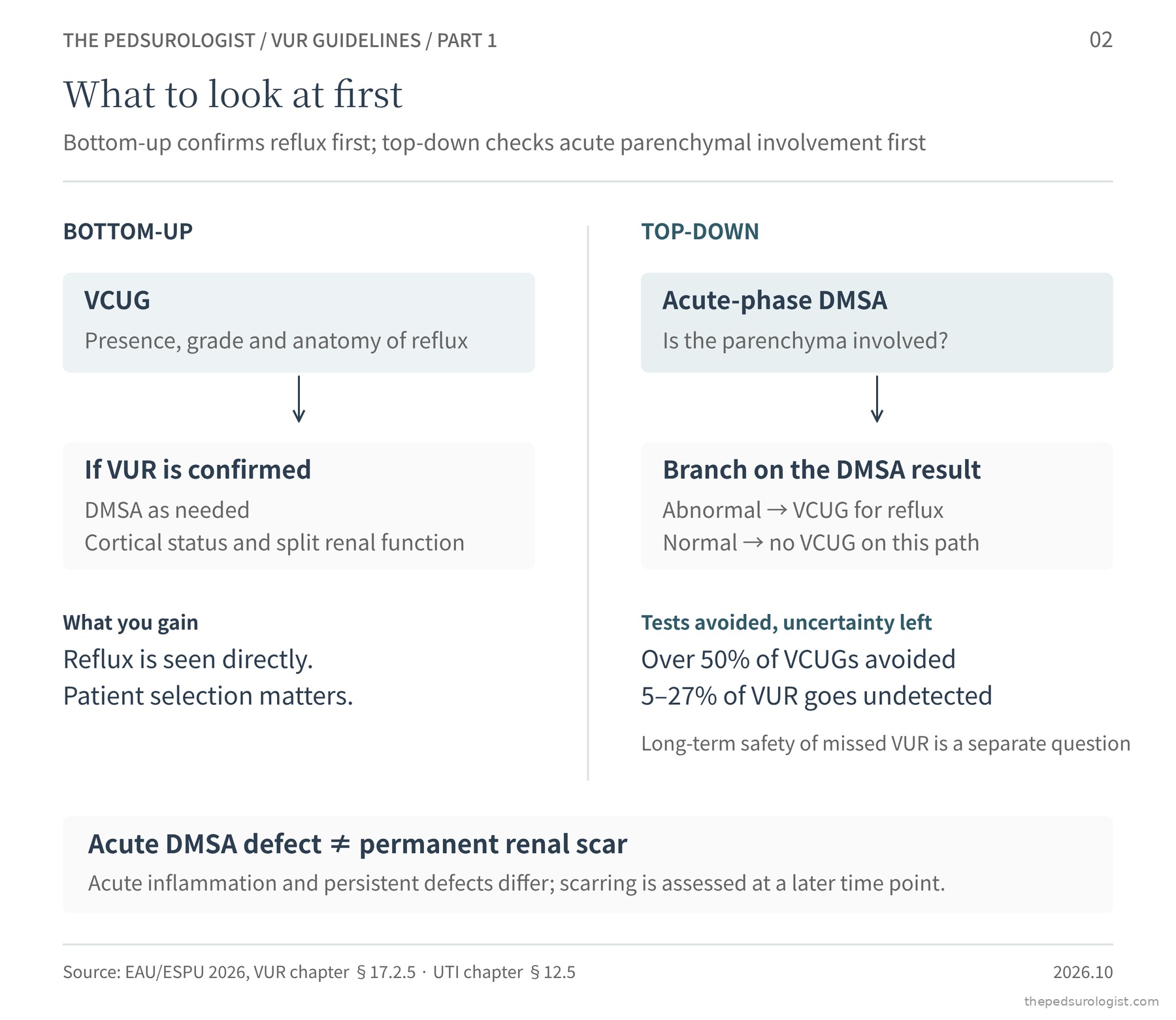
Top-down is an alternative that EAU/ESPU describes in its text. A DMSA scan is performed first, during the acute infection, and VCUG only if the parenchyma is involved. It avoids more than half of VCUGs at the cost of missing 5–27% of reflux. That the missed reflux is less important is an assumption the guideline itself qualifies with "presumably", and the long-term safety of the strategy has not been established. An acute-phase DMSA defect cannot by itself be taken as a permanent scar: acute inflammation may be mixed with pre-existing scarring or parenchymal abnormality, and scarring is judged six months later.
3. Why does an atypical UTI justify imaging?
All three guidelines treat an "atypical" infection as an indication for imaging, but they define it differently. EAU/ESPU describes it by contrast with a typical infection (common organism, response to oral antibiotics within 48 hours, no need for admission); NICE lists criteria; and the AAP, rather than using an "atypical" category, names the situations in which VCUG may be chosen even after a first infection. Laid out item by item, the overlap becomes visible.
| Item | EAU/ESPU | NICE NG224 | AAP 2026 |
|---|---|---|---|
| Non-E. coli organism | ● | ● | ● |
| Sepsis / severe course | ● Course that may need admission | ● Seriously ill, septicaemia | ● Septic shock, ICU admission |
| No response within 48 h | ● Fails or cannot tolerate oral treatment | ● | — |
| Multidrug-resistant organism | ● | — | — |
| Poor urine flow | — | ● | — |
| Abdominal or bladder mass | — | ● | — |
| Raised creatinine | — | ● | — |
| Age under 1 month | — | — | ● |
| Circumcised boy | — | — | ● |
| Follow-up uncertain | — | — | ● |
Only two items appear in all three: infection with an organism other than E. coli, and a severe course. Slow response to treatment is shared by the two European guidelines. The rest differ. NICE adds clinical and laboratory findings that suggest obstruction or impaired renal function; the AAP adds groups in which infection is rare enough that a single episode carries more weight (neonates, circumcised boys) and the practicalities of follow-up.
These findings are treated as indications because they may signal an underlying abnormality of the urinary tract. EAU/ESPU notes that a UTI may be the first presentation of a previously unknown anomaly (posterior urethral valves, stricture, stones, lower urinary tract dysfunction, reflux), and explains that complicated UTI usually arises as an ascending infection on top of pre-existing uropathy such as reflux or obstruction.
Why a non-E. coli organism should be such a signal is classically explained by differences in virulence. Uropathogenic E. coli can ascend a normal urinary tract using adhesins such as P fimbriae; if an organism lacking that equipment causes pyelonephritis, one first suspects that conditions favouring ascent — reflux, obstruction, stasis — were already present. What the guidelines actually present, however, is not the mechanism but an association between non-E. coli UTI and urinary tract abnormality. In a Finnish series of 134 children with bacteraemic UTI, obstruction or reflux was present in 46% of E. coli infections and 89% of non-E. coli infections — figures from a selected, bacteraemic population9. The AAP likewise names a non-E. coli organism as the risk factor most consistently associated with reflux in children with UTI.
A severe course follows a different logic. Sepsis, or a course that needs admission, in itself raises the likelihood of parenchymal involvement and bacteraemia, and delayed treatment increases the risk of scarring. A severe course is therefore a reason to assess both the extent of parenchymal involvement and causes such as obstruction, bringing ultrasound forward to the acute phase and continuing through to assessment of scarring. The AAP's inclusion of circumcised boys has yet another rationale: infection is so uncommon in that group that a single episode means more.
The guidelines do not explain what each remaining item points to. The clinical reading is that poor urine flow or a bladder mass first raises bladder outlet obstruction, a raised creatinine the possibility of acute or pre-existing renal impairment, and failure to respond within 48 hours obstruction or abscess. These are not measured predictive values for each item.
4. Should siblings and infants with prenatal hydronephrosis be screened?
The figures both guidelines quote come from the same meta-analysis. Reflux was found in 27.4% of screened siblings and 35.7% of offspring, and renal damage in 14.4% of asymptomatic siblings. These are estimates from screened research populations, with wide ranges. Reflux found by screening tends to be low grade and to resolve early. A high prevalence and evidence that screening improves that child's outcome are two separate things. There is no randomised trial comparing screened and unscreened groups.
EAU/ESPU's recommendation is therefore not to screen but to inform: tell parents that siblings and offspring have a high prevalence of reflux (Strong). The AUA recommends cystography in a sibling with renal cortical abnormalities or size asymmetry on ultrasound, or a history of UTI (Recommendation), and leaves all other screening as an Option. The reason is the same: the health benefit of finding reflux has not been demonstrated.
Infants with prenatal hydronephrosis are similar. Reflux prevalence among screened infants is 16.2%, and the degree of hydronephrosis does not predict it. The AUA recommends VCUG for SFU grade 3–4 hydronephrosis, hydroureter, an abnormal bladder, or a UTI under observation (Recommendation), and leaves both observation and VCUG as options for grades 1–2. EAU/ESPU regards reflux as uncommon when two ultrasounds in the first one to two months are both normal, and advises VCUG for bilateral high-grade hydronephrosis, duplex kidneys with hydronephrosis, ureterocele, ureteric dilatation and abnormal bladders (narrative text).
Both guidelines end in the same place: do not test everyone, but make sure the family knows not to delay a urine test when the child has a fever.
Closing Part 1
Whether reflux found later by selective testing changes the prognosis, and whether finding it in asymptomatic siblings reduces infection and renal damage, have never been measured directly. That testing can find reflux, and that finding it improves the child's prognosis, are different questions. The guidelines are moving towards selective testing because the first question can be answered while the second still lacks a sufficient answer. Part 2 looks at how children with confirmed reflux are stratified and followed.
A note on currency
- The 2026 EAU/ESPU edition is a limited update from March 2026. The UTI chapter was revised in this edition; the VUR chapter reflects its 2023 revision (Gnech 2024).
- The imaging recommendations of NICE NG224 (2022) are carried over unchanged from CG54 (2007).
- The AAP's 2011 UTI guideline was retired in 2021 after criticism of the use of race as a variable in its urine-sampling risk calculator; a new guideline for children aged 8 days to 5 years appeared in September 2026.
- The AUA guideline was published in 2010 and amended in 2017.
Guideline texts checked on 19 October 2026.
References
- EAU Guidelines on Paediatric Urology. Chapter 17, Vesicoureteric reflux; Chapter 12, Urinary tract infections in children. European Association of Urology, 2026 edition (limited update March 2026). uroweb.org
- Gnech M, 't Hoen L, Zachou A, et al. Update and summary of the European Association of Urology/European Society of Paediatric Urology paediatric guidelines on vesicoureteral reflux in children. Eur Urol. 2024;85(5):433-442. doi:10.1016/j.eururo.2023.12.005
- Peters CA, Skoog SJ, Arant BS Jr, et al. Summary of the AUA guideline on management of primary vesicoureteral reflux in children. J Urol. 2010;184(3):1134-1144. doi:10.1016/j.juro.2010.05.065
- Skoog SJ, Peters CA, Arant BS Jr, et al. Pediatric vesicoureteral reflux guidelines panel summary report: clinical practice guidelines for screening siblings of children with vesicoureteral reflux and neonates/infants with prenatal hydronephrosis. J Urol. 2010;184(3):1145-1151. doi:10.1016/j.juro.2010.05.066
- American Urological Association. Management and screening of primary vesicoureteral reflux in children (2010, amended 2017). auanet.org
- National Institute for Health and Care Excellence. Urinary tract infection in under 16s: diagnosis and management. NICE guideline NG224, 2022 (imaging recommendations from CG54, 2007). nice.org.uk/guidance/ng224
- Alverson BK, Hains DS, Downs SM, et al. Clinical practice guideline for the diagnosis and treatment of urinary tract infection in children from 8 days to 5 years of age. Pediatrics. 2026;158(4):e2026078565. publications.aap.org
- Subcommittee on Urinary Tract Infection; Roberts KB. Urinary tract infection: clinical practice guideline for the diagnosis and management of the initial UTI in febrile infants and children 2 to 24 months. Pediatrics. 2011;128(3):595-610. doi:10.1542/peds.2011-1330 (retired 2021)
- Honkinen O, Jahnukainen T, Mertsola J, Eskola J, Ruuskanen O. Bacteremic urinary tract infection in children. Pediatr Infect Dis J. 2000;19(7):630-634. doi:10.1097/00006454-200007000-00009