
Few topics in paediatric urology have generated so much literature and so little agreement. More than a century after Keyes first described it in 1919, concealed penis still has no internationally settled terminology, no accepted classification, and no defined surgical indication. In Korea, a blanket reimbursement denial for penoplasty some years ago prompted a society-level response — and that dispute, too, traces back to the same root. Without a defined indication, the same operation is a functional correction to one reviewer and a cosmetic procedure to another.
This is written in two parts. Part 1 covers terminology, aetiology, classification, and the under-discussed question of penile size. Indications, timing and surgical technique follow in Part 2.
Terminology — why it never converged
Keyes described "an apparent absence of the penis which exists when the penis lacks its proper sheath of skin, lies buried beneath the integument of the abdomen, thigh or scrotum." Since then, buried penis, concealed penis, inconspicuous penis, hidden penis, congenital megaprepuce, trapped penis and webbed penis have been used interchangeably, each author applying their own criteria. Korean practice is no different: several vernacular terms coexist and mean different things to different clinicians.
There are two reasons the terminology has not settled. First, there is no threshold for how much loss of exposure counts as abnormal. Normal exposure itself has never been defined, so no boundary can be drawn. Second, this is a spectrum disorder — the appearance varies from child to child according to the severity of each contributing factor and the size of the penis.
Hadidi (2014) offered a concrete proposal to end the confusion: reserve buried penis for the congenital spectrum and use concealed penis only for acquired presentations later in life due to fat accumulation. It is logically tidy but does not match how the terms are used in Korean clinics. This article keeps concealed penis as the umbrella term, in line with established usage, while noting that the proposal exists. That the choice of word is itself still contested tells you where the subject stands.
Aetiology — observations that contradict each other
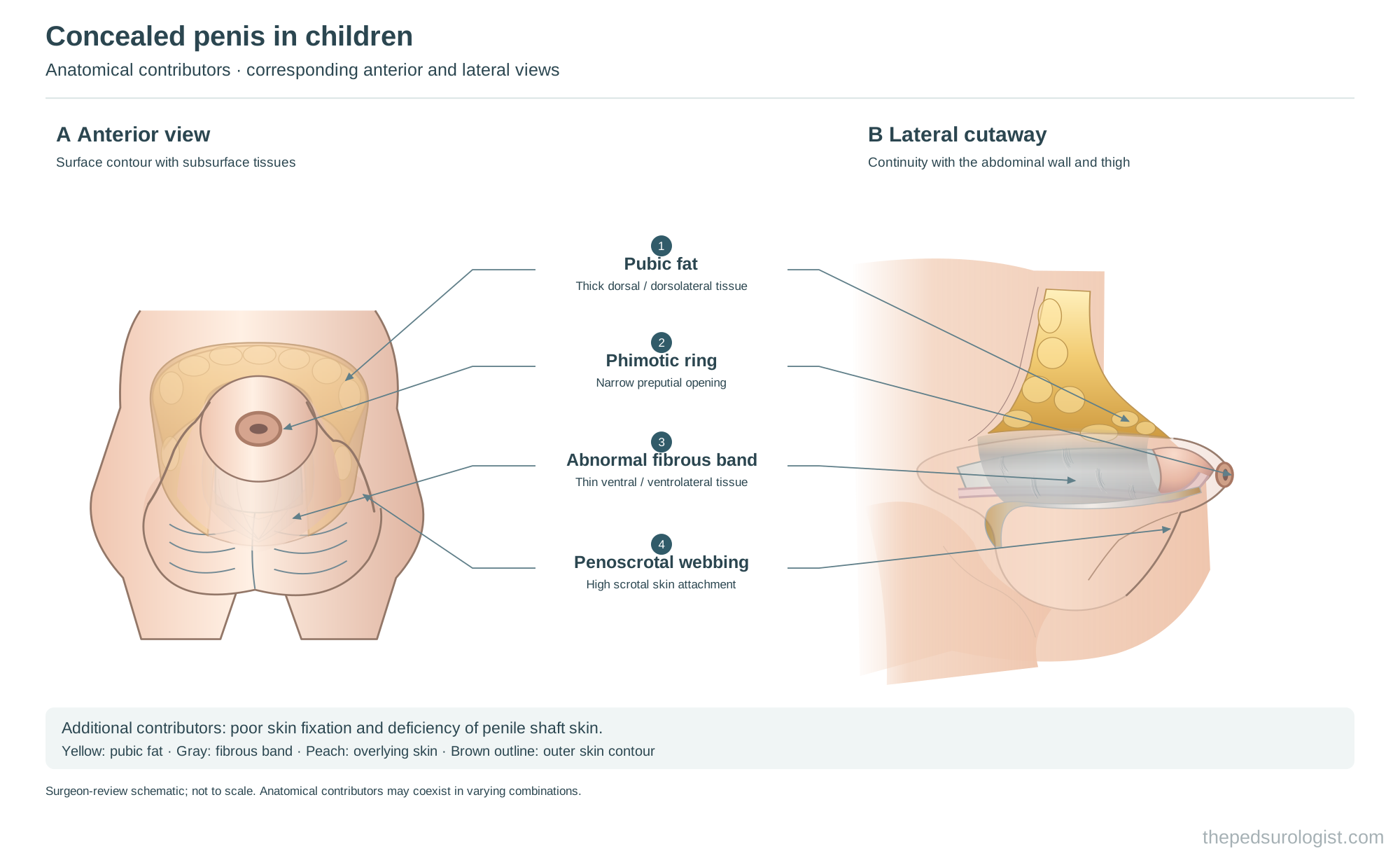
The most widely accepted primary cause is an abnormal fibrous band: a circumferential dysplastic cord running from Scarpa's fascia above the pubis down beneath the penis to the scrotum, tethering the shaft inward. This band has also been described as the distal attachment of one of the penile suspensory structures, the fundiform ligament. To it are added an absolute deficiency of penile skin, abnormal attachment between shaft and skin, and penoscrotal fusion.
Yet the published observations disagree substantially. Crawford described a dartos-like fibromuscular layer tethering the shaft to the abdominal wall, whose division allowed the penis to assume its normal relationship. Wollin et al. emphasised abnormal mobility of the skin over the shaft. Hadidi found abnormal mobility of skin and fascia in all 61 of his patients and abnormal attachment of the fundiform and suspensory ligaments in 32. Redman, by contrast, observed no abnormality of the tunica dartos and no tethering bands in any of his 31 personal cases.
When one surgeon reports a finding as constant and another reports it as absent in the same disease, either they are looking at different things or they are looking at different diseases. The latter is more likely, and it is one reason no classification has held.
The overlooked axis — ratio, not fat
Exposure is also commonly impaired by excess suprapubic fat with no morphological abnormality at all. What is often missed here is that the judgement "fat is hiding the penis" is always relative to penile size. The same fat pad is no problem over a larger penis.
By definition, concealed penis excludes micropenis. In practice, however, a substantial number of patients fall into a gray zone: smaller than normal, but not below the −2.5 SD threshold that defines micropenis. Converting −2.5 SD to a percentile gives roughly the bottom 0.5% — which leaves a wide band above it.
The gray zone goes unrecognised because it is hard to measure. Penile size in children is assessed by stretched penile length, and in a penis that is difficult to expose and difficult to stretch, an accurate measurement is not easily obtained.
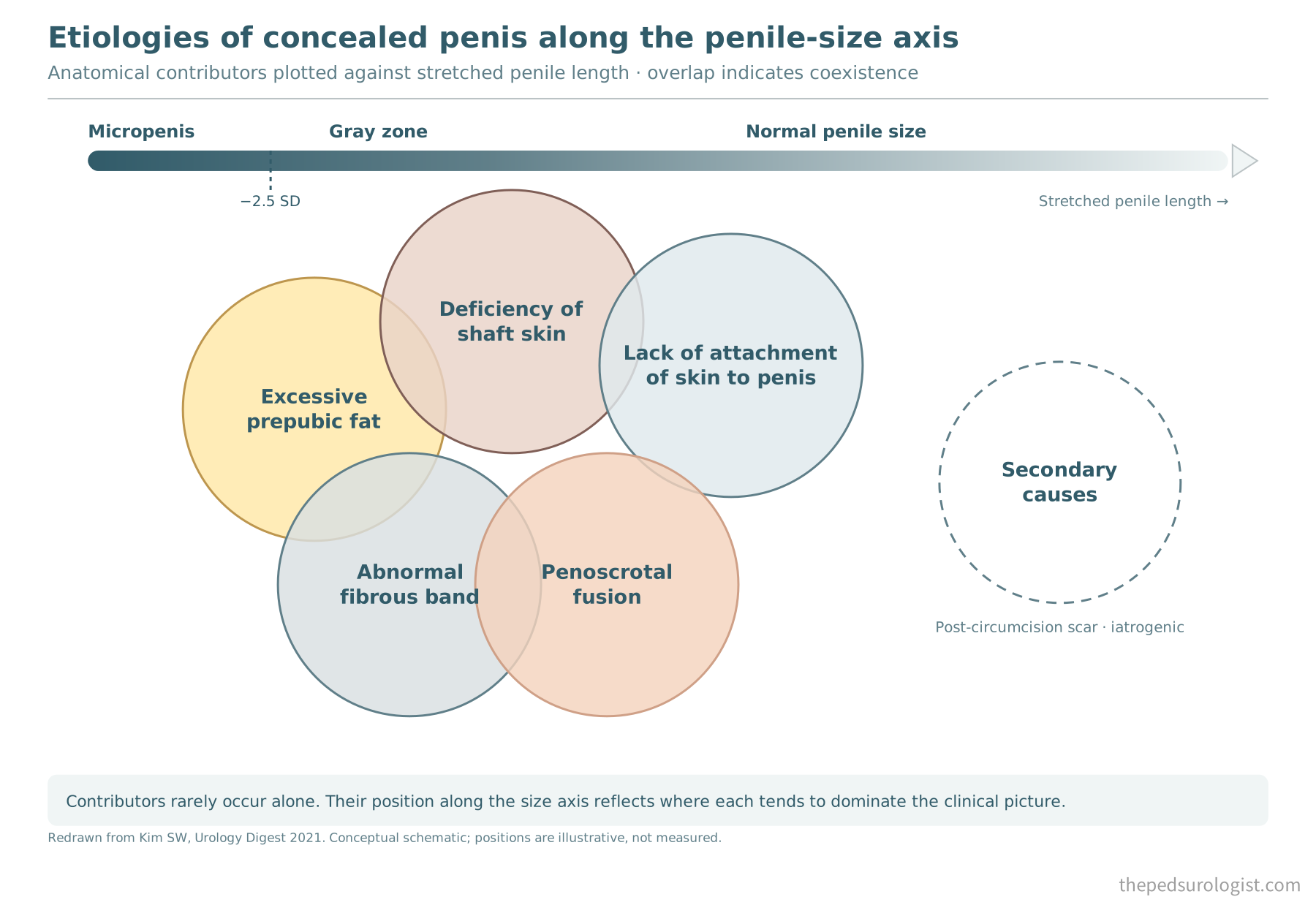
The diagram above, first presented in my 2021 review, shows the relationship at a glance. The horizontal axis is penile size; each anatomical contributor sits in the range where it tends to dominate the clinical picture. Excess prepubic fat straddles the gray zone; lack of skin attachment sits toward normal size. The same contributor may or may not be a problem depending on penile size — that is the point of the layout — and the overlaps reflect the fact that in real patients these factors coexist.
Even so, the measurements already exist in the literature. In Hadidi's 61 patients (mean age 15 months) the mean stretched penile length was 3.3 cm (range 2.9–4.5), and 47 had a short penile shaft. Redman reported a mean of 3.1 cm in 31 boys (mean age 12 months). The normative values cited in the same paper, from Elder, are 4.3 ± 0.8 cm at 6–12 months and 4.7 ± 0.8 cm at 1–2 years.
This population is, on average, smaller than normal. Yet Donahoe and Radhakrishnan reported that corporeal and glanular size and development are normal in buried penis and that the penis is of normal size — and textbook descriptions still largely follow that line. What is striking is that Hadidi and Redman themselves, having reported below-average measurements, did not draw from them the conclusion that penile size is one axis of the disease. The measurement has been made; the interpretation has not. The gray-zone argument does not need new data — it needs the existing data to be read properly.
Classification — a history of attempts
The first systematic attempt was Crawford's in 1977, dividing cases by degree of exposure into concealed penis, buried penis (partial or complete) and penoscrotal web. In 1986 Maizels et al. proposed four categories by mechanism of concealment — buried, webbed, trapped and micropenis — with technique chosen by category.
The 2015 review by Cimador et al. in Nature Reviews Urology placed concealed penis as the parent term with buried, webbed and trapped penis beneath it, borrowing earlier definitions of buried penis without clarifying them.
One finding has drawn attention across all these classifications: an abnormality of the inner prepuce and the dartos beneath it — an inner prepuce that is excessively long over a dartos that is thick and inelastic. In severe cases urine is trapped within the prepuce during voiding, the genital area balloons, and the urine must be expressed by hand. Authors have given this different names, but the entity is the same.
How this dartos actually differs has been examined histologically. In a prospective comparison of dartos from 20 buried penis, 20 hypospadias and 20 normal cases (Shalmont et al., 2018), the buried-penis dartos showed collagen fibres that were thicker but fewer, with reduced elastin — a pattern resembling the dartos in hypospadias. Fragmented elastin has also been reported in buried-penis dartos. In the same study, however, the tissue was well vascularised on CD31 staining, and the authors concluded it was not fibrotic in character. A thick, inelastic dartos is a common accompaniment of concealed penis, and it histologically resembles that of hypospadias — that is the picture to date.
The finding matters clinically for two reasons. One is postoperative oedema. Tissue with reduced and fragmented elastin has poor compliance and may drain lymph poorly; add surgical dissection and oedema forms easily and persists. The other is the shape that the oedema leaves behind: while the thick dartos is swollen the penile contour is blunted, and after the swelling settles the skin can appear redundant. This remains a hypothesis rather than a verified mechanism, but it is a candidate explanation for why oedema and the resulting dissatisfaction with appearance are so common after this operation, and it is taken up again in Part 2.
Hadidi (2014) made this inner-preputial abnormality (which he calls long inner prepuce, LIP) the axis of his classification: the abnormality alone is Grade I, with abnormal attachment of the fundiform and suspensory ligaments Grade II, and with excess suprapubic fat Grade III.
He also argues that the ballooning presentation (named congenital megaprepuce by O'Brien et al. in 1994) is not a separate entity but a variant of Grade I. Re-examining those cases, he holds that the excess is in the inner prepuce and that the intermittent ballooning is due to a valve-like mechanism rather than phimosis. Shenoy and Ferro take the opposite view, treating it as a distinct entity requiring a different surgical plan.
What I actually look at
The existing classifications are ambiguous for the reasons above. When I assess a patient, three axes matter.
First, the ratio of penile size to surrounding tissue. This is broader than "suprapubic fat," and it is ultimately what determines exposure. The gray-zone problem sits here.
Second, the presence of penoscrotal fusion (webbing). A structural problem that growth does not resolve.
Third, the presence of an inner-dartos abnormality. Being an internal finding it is hard to predict before surgery, but it can influence the postoperative result and is worth bearing in mind.
I use these three not because they are a more accurate classification but because each leads to a different management decision. If the first axis dominates, surgery has little to offer; the second is corrected only by surgery; the third changes the choice of technique.
That is the diagnostic and conceptual ground. How these three axes are read determines whether to operate and how — the subject of Part 2.
References
- Keyes EL Jr. Phimosis, paraphimosis, tumors of the penis. Urology. New York: Appleton; 1919. p. 649.
- Crawford BS. Buried penis. Br J Plast Surg. 1977;30(1):96-99.
- Maizels M, Zaontz M, Donovan J, Bushnick PN, Firlit CF. Surgical correction of the buried penis: description of a classification system and a technique to correct the disorder. J Urol. 1986;136(1):268-271.
- O'Brien A, Shapiro AMJ, Frank JD. Phimosis or congenital megaprepuce? Br J Urol. 1994;73(6):719-720.
- Cromie WJ, Ritchey ML, Smith RC, Zagaja GP. Anatomical alignment for the correction of buried penis. J Urol. 1998;160(4):1482-1484.
- Redman JF. Buried penis: congenital syndrome of a short penile shaft and a paucity of penile shaft skin. J Urol. 2005;173(5):1714-1717.
- Hadidi AT. Buried penis: classification surgical approach. J Pediatr Surg. 2014;49(2):374-379.
- Shalmont G, Situmorang GR, Wahyudi I, Tanurahardja B, Rodjani A, Atmoko W. Abnormal dartos fascia in buried penis and hypospadias: evidence from histopathology. J Pediatr Urol. 2018;14(6):536.e1-536.e7.
- Cimador M, Catalano P, Ortolano R, Giuffrè M. The inconspicuous penis in children. Nat Rev Urol. 2015;12(4):205-215.
- Kim SW. Concealed penis from the perspective of pediatric urology. Urology Digest. 2021;2(2). (in Korean)