
Part 1 (The Shifting View of Pathogenesis and Prognosis) covered how our understanding of why cryptorchidism occurs has changed, and the long-term prognosis by paternity rate. Part 2 covers how the actual surgical technique is chosen by position and situation.
Approach Selection — Scrotal vs. Inguinal
For a palpable undescended testis, choosing between a scrotal approach and the traditional inguinal approach is a practical question. The scrotal approach, first described by Bianchi and Squire in 1989, is easy to apply through a single incision for less severe positions — a high scrotal or low inguinal testis — with a shorter operative time.
Its limits are just as clear, though. A scrotal incision alone makes it structurally difficult to dissect far enough to reach the area near the internal ring — the technique works by retracting the external ring upward and inspecting the spermatic cord structures through that window, which limits the field of view and range of manipulation any further proximally. This makes it harder to directly visualize and dissect the proximal testicular vessels, and early series already reported that the scrotal approach fails and converts to an inguinal incision more often the closer the testis sits to the high inguinal region or the internal ring (Bianchi & Squire, 1989; Bassel et al.). Recent meta-analyses report a conversion rate from scrotal to inguinal of roughly 3.6-7.3%.
In practice, then, it is safer to treat the scrotal approach as an option to consider only when the testis is palpable down around the upper scrotum. When the position is uncertain or judged to be close to the internal ring, starting with an inguinal approach is the reasonable choice — and even when beginning scrotally, the possibility of converting to an inguinal incision should always be kept open. Laparoscopy was originally introduced for diagnosing and treating a non-palpable testis; applying it uniformly even to palpable, low-lying testes is better understood as operator or institutional preference than as standard technique (see the callout below).
When Is Laparoscopy Indicated?
Laparoscopy's original indications are twofold. First, localizing a non-palpable testis — when nothing is felt in the inguinal region or abdomen, the poor diagnostic performance of imaging makes laparoscopy the de facto reference standard. A meta-analysis pooling ultrasound's diagnostic performance for the non-palpable testis (12 studies, 591 testes) found only 45% sensitivity and 78% specificity (Tasian & Copp, 2011) — meaning a negative result can't reliably be used to rule out an intra-abdominal testis. Second, treating a high intra-abdominal testis — when the spermatic vessels are too short to bring the testis all the way to the scrotum in one operation, a staged approach is needed: initial vessel management via laparoscopy followed by a second-stage operation.
Single-Stage vs. Two-Stage Surgery — How Atrophy Rates Changed Over Time
Outcomes for the high intra-abdominal testis have followed a clear trend over the past several decades. In the open-surgery era, Fowler-Stephens atrophy rates were reported to be considerably high; with the introduction of laparoscopy, more precise visualization and minimally invasive dissection became possible, and atrophy rates improved dramatically — an improvement that's especially pronounced when combined with a two-stage (staged) approach. The approximate ranges reported in the literature are summarized below.
| Technique | Reported atrophy rate |
|---|---|
| Standard (low) orchiopexy | ~0.15-3% (pooled across cohorts) |
| Fowler-Stephens, single-stage | ~17.3% (pooled, 2025 meta-analysis) |
| Fowler-Stephens, two-stage | ~11% (pooled, 2025 meta-analysis) |
The idea of staging itself arose as a response to these atrophy rates. The technique Fowler and Stephens described in 1959 was a single-stage operation, dividing the vessels and moving the testis in one sitting. In 1984, Ransley et al. proposed ligating the testicular vessels at a first operation without touching the testis itself at all, allowing time for collateral supply along the vas deferens to develop, and only then mobilizing and bringing the testis down at a second operation. Staged approaches became standard thereafter.
The most recent systematic review and meta-analysis (Fung et al., 2025) confirmed this contrast was statistically significant — the two-stage approach, compared with single-stage, significantly improved overall success rate (OR 2.57, 95% CI 1.50-4.39, p=0.0006) and significantly lowered atrophy risk (OR 0.48, 95% CI 0.28-0.79, p=0.004). This is explained by the two-stage approach giving collateral vessels time to develop sufficiently.
Is Diagnostic Laparoscopy Necessary?
Whether diagnostic laparoscopy can be skipped for a non-palpable testis is a practically important question, and the answer generally leans toward "necessary." A testis that remains non-palpable even under exam under anesthesia is one of three things: ① vessels and vas deferens ending blindly (vanishing testis), ② an intra-abdominal testis, or ③ a testicular nubbin or atrophied testicular remnant — and no method distinguishes among these three as accurately, or leads as directly to treatment, as laparoscopy. Attempts to confirm an intra-abdominal testis beforehand with ultrasound or MRI suffer from low sensitivity, making a negative result unreliable for ruling anything out.
The Shehata Technique — A Complementary Alternative
The fundamental trade-off of the Fowler-Stephens procedure is that it sacrifices the spermatic vessels in favor of relying on collaterals. The Shehata (traction) technique, increasingly used in recent years, takes a different approach to this same premise. Rather than ligating the spermatic vessels, the testis is sutured to the abdominal wall at the first operation, applying traction that gradually lengthens the spermatic cord; a few weeks to roughly 3 months later, the second operation brings the testis down to the scrotum with the vessels preserved.
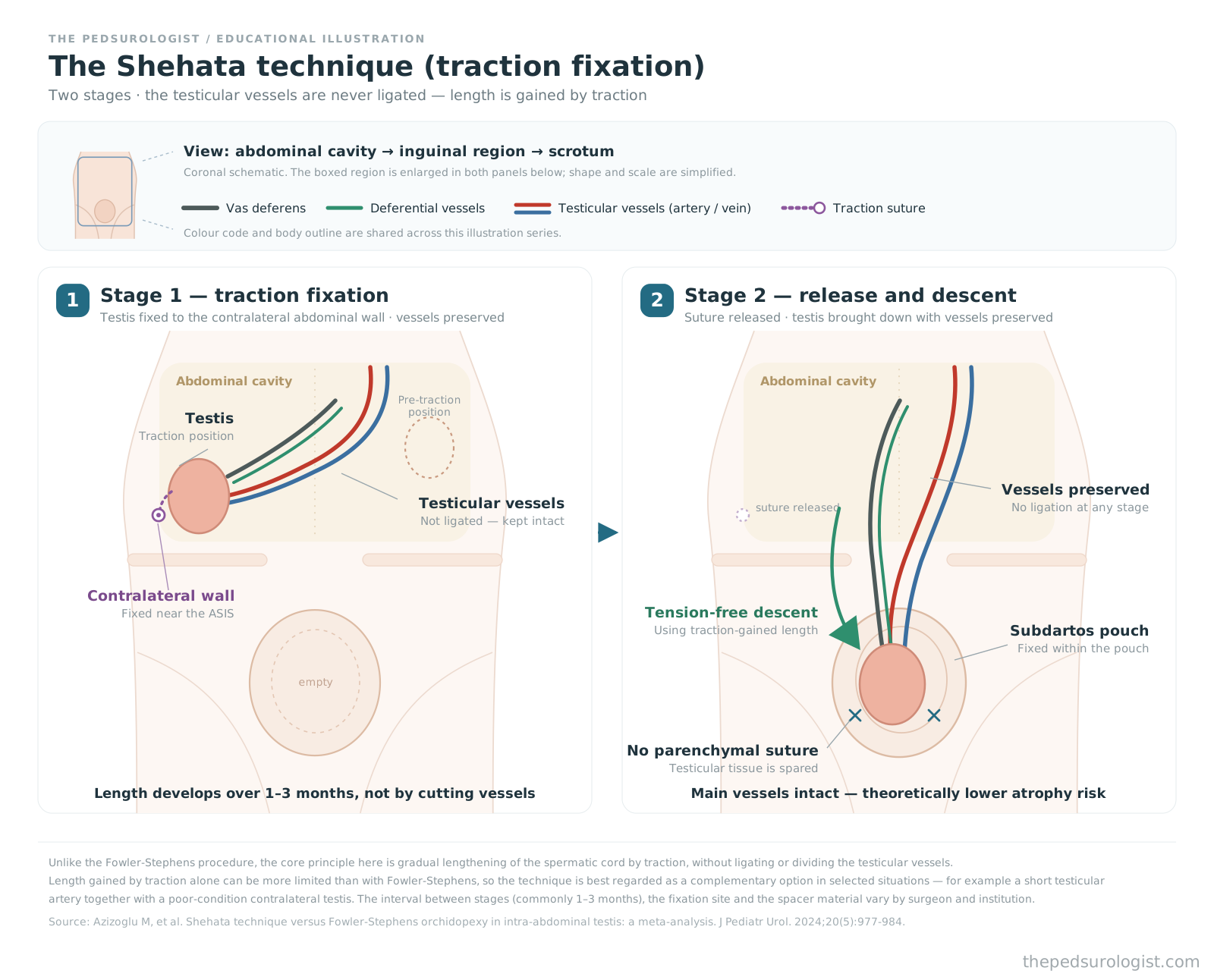
Per a meta-analysis (Azizoglu et al., 2024, J Pediatr Urol), the Shehata technique showed a higher overall success rate (87% vs. 74%) and a lower atrophy rate (5% vs. 12%) compared with Fowler-Stephens. The underlying rationale for preserving the main vessels is clearly favorable for preventing atrophy in principle.
That said, from personal surgical experience, I'll add one caveat: achieving as much descent length with the Shehata technique as with Fowler-Stephens hasn't always been straightforward. There's a practical ceiling to how much length traction alone can secure. Rather than replacing the entire category of high intra-abdominal testes with this technique, it's closer to practice to think of it as a complementary technique for specific conditions — for instance, when the testicular artery is somewhat short, making conventional orchiopexy (the standard technique that preserves and brings down the vessels, not Fowler-Stephens) precariously difficult, and at the same time the contralateral testis isn't in good condition, so avoiding atrophy is a priority. Being a relatively recent technique, it's still performed at a limited number of centers domestically, and long-term fertility data haven't accumulated to the same extent as for Fowler-Stephens.
I've increasingly encountered cases in clinical practice where laparoscopic orchiopexy is applied uniformly even to extra-abdominal (inguinal, high-scrotal) testes. My personal concern is that, in some cases, this trend appears to be driven more by reimbursement and marketing incentives than by clinical evidence. Laparoscopy was developed for non-palpable and intra-abdominal testes. When applied aggressively to an inguinal testis, excessive dissection around the internal ring to gain sufficient length risks damaging normal structures, and the traction process of pulling the testis up into the abdomen and repositioning it may actually raise the risk of injury to the testicular vessels and vas deferens. I want to be clear that this reflects a pattern I've personally observed in clinical practice rather than an academically established statistic, and it is not a conclusion supported by the literature.
References
- Bianchi A, Squire BR. Transscrotal orchidopexy: orchidopexy revised. Pediatr Surg Int. 1989;4(3):189-192.
- Bassel YS, Scherz HC, Kirsch AJ. Scrotal incision orchiopexy for undescended testes with or without a patent processus vaginalis. J Urol. 2007;177:1516-1518.
- Tasian GE, Copp HL. Diagnostic performance of ultrasound in nonpalpable cryptorchidism: a systematic review and meta-analysis. Pediatrics. 2011;127(1):119-128.
- Fung ACH, Tsang JTW, Leung L, Chan IHY, Wong KKY. Comparative Outcomes of Single-Stage versus Two-Stage Laparoscopic Fowler-Stephens Orchidopexy: A Systematic Review and Meta-Analysis. Eur J Pediatr Surg. 2025;35(1):28-35.
- Fowler R, Stephens FD. The role of testicular vascular anatomy in the salvage of high undescended testes. Aust N Z J Surg. 1959;29(1):92-106.
- Ransley PG, Vordermark JS, Caldamone AA, Bellinger MF. Preliminary ligation of the gonadal vessels prior to orchidopexy for the intra-abdominal testicle: a staged Fowler-Stephens procedure. World J Urol. 1984;2(4):266-268.
- Bidault-Jourdainne V, et al. Staged laparoscopic orchiopexy of intra-abdominal testis: spermatic vessels division versus traction? A multicentric comparative study. J Pediatr Urol. 2024.
- Azizoglu M, Zain M, Karakas E, Kotb M, Kamci TO, Bakir AC, Shehata S. Shehata technique versus Fowler-Stephens orchidopexy in intra-abdominal testis: a meta-analysis. J Pediatr Urol. 2024;20(5):977-984.