Urinary tract dilation detected on prenatal ultrasound is not the picture of a single disease. In some children only the renal pelvis and calyces dilate; in others the ureter dilates as well. Even dilation that looks similar can follow different courses.
Part 1 looked at what we measure and how we grade it, and Part 2 at which tests to perform in which children after birth. This time we go back to an earlier point. How does the ureter begin, how does it connect to the bladder, and from when does it become a structure that transports urine?
Same hydronephrosis, so why does it dilate in different places
Development of the urinary tract involves several tasks that proceed in overlapping fashion. A collecting system that gathers urine must be formed, the ureter must connect to the bladder, and the musculature and contractile function of the ureteral wall must mature. Depending on which process goes wrong, similar findings of dilation can carry different meanings.
Hydronephrosis describes a form, namely dilation. Embryology explains the background against which that form arises, but dilation alone cannot establish current obstruction or the need for surgery.
The ureteric bud and renal tissue develop together
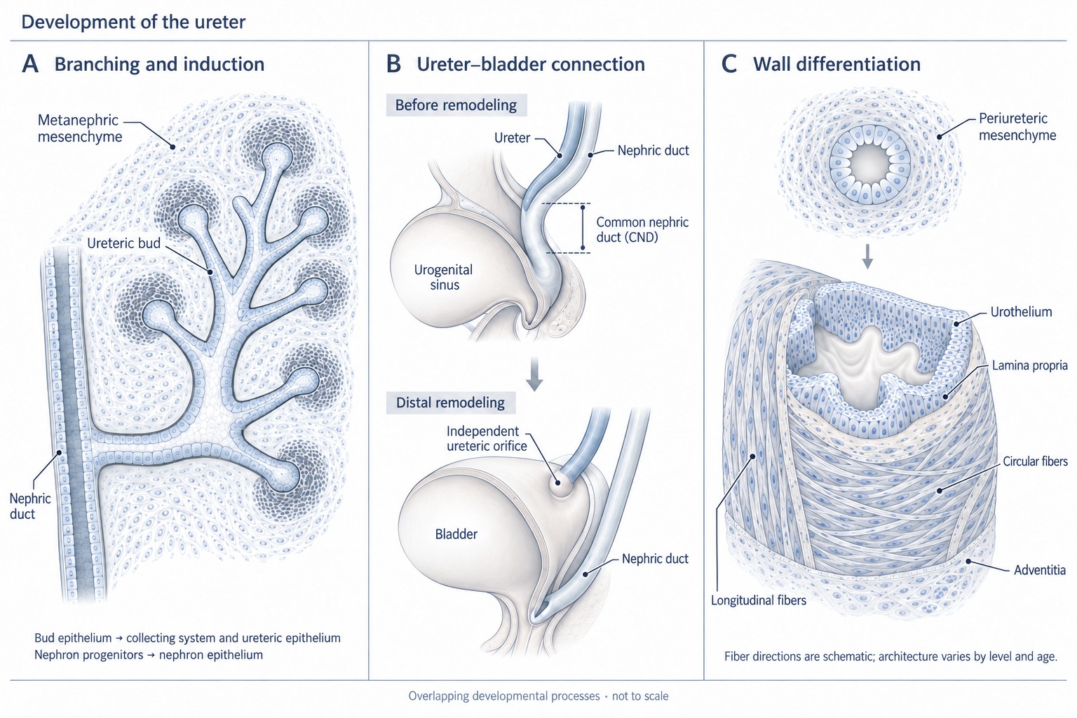
The ureter starts as the ureteric bud, which grows out from the mesonephric duct. The ureteric bud enters the metanephric mesenchyme and branches, and the two tissues develop through reciprocal signaling. GDNF secreted by the metanephric mesenchyme and the RET receptor on the ureteric bud are the representative signaling system involved in this interaction. In mice lacking RET or GDNF, ureteric bud formation and branching were absent or incomplete, and the kidney either failed to form or showed severe developmental abnormalities (Schuchardt et al., 1994; Pichel et al., 1996).
It helps here to separate the origins of the structures. The epithelium of the ureteric bud gives rise to the epithelium of the ureter, renal pelvis, calyces and collecting ducts. The main epithelial components of the nephron, by contrast, are formed from nephron progenitor cells of the metanephric mesenchyme. The smooth muscle and supporting tissue of the ureteral wall depend on differentiation of the mesenchyme surrounding the epithelium. The ureteral wall as a whole is not derived from ureteric bud epithelium (Airik et al., 2006).
This distinction matters clinically as well. The collecting system and the renal parenchyma are not structures completed separately and then joined, but tissues that influence each other during development. Ureteral abnormalities and abnormal development of the renal parenchyma can therefore occur together. That said, not all renal dysplasia can be explained as a consequence of ureteral obstruction. In a study analyzing 51 duplex kidneys, the degree of renal dysplasia was more closely associated with how far the ureteral orifice of that moiety was displaced from its normal position than with the presence of obstruction (Mackie and Stephens, 1975).
Scroll horizontally to see the full figure
The ureter separates from the mesonephric duct and connects to the bladder
The early ureteric bud branches off the mesonephric duct. The ureter must then change this connection and form an independent opening into the bladder. The segment of the mesonephric duct lying between the origin of the ureteric bud and the cloaca (urogenital sinus) at this stage is called the common nephric duct (CND).
The classic scheme holds that the CND is incorporated into the bladder to become the trigone, and that the ureteral orifice moves as this happens. Mouse studies showed instead that the CND does not differentiate into the trigone but disappears by apoptosis, and that this process is the key step that detaches the ureter from the mesonephric duct and connects it to the bladder (Batourina et al., 2005). In mouse lineage-tracing studies, contrary to the earlier model, the trigone was formed mainly from bladder smooth muscle, with only a small ureteral contribution, and arose as ureteral and bladder muscle interlocked (Viana et al., 2007). The change in ureteral position can therefore be understood as a process combining removal of distal tissue with formation of a new connection.
The importance of this process is also seen in animal models with abnormal maturation of the distal ureter. In mice with combined deficiency of the LAR family receptor protein tyrosine phosphatases (Ptprs and Ptprf), reduced programmed cell death in the distal ureter led to hydroureter and ureterocele (Uetani et al., 2009). These experiments, however, are evidence for the importance of a particular developmental process; they do not explain every human ureterocele or distal obstruction by a single cause.
Early branching of a single ureteric bud and formation of two buds are the classic developmental models for incomplete and complete duplication. In complete duplication, the Weigert–Meyer rule, by which the upper-moiety ureter opens below and medial to the lower-moiety ureter, commonly holds. The upper moiety is associated with an ectopic orifice and ureterocele, and the lower moiety with reflux, but there are exceptions. The further the ureteral orifice was displaced from its normal position, the more severe the hypoplasia and dysplasia of that moiety (Mackie and Stephens, 1975). Duplication in itself does not mean obstruction.
For an open passage to transport urine
The ureter is not a passive tube down which urine flows by gravity. Urine is carried to the bladder only when contractions initiated in the renal pelvis propagate in a coordinated way along the ureter. This requires not only smooth muscle differentiation but also fascicle arrangement, intercellular connections, and a system that regulates and propagates contraction. Interstitial cells of Cajal are described as cells involved in this pacemaking and peristalsis (Babu et al., 2019).
In mice lacking Tbx18, which regulates development of the ureteral mesenchyme, ureteral mesenchymal cells failed to differentiate into smooth muscle and instead became fibrous, ligament-like tissue, and a short hydroureter and hydronephrosis were present at birth (Airik et al., 2006). The result shows that development of the ureteral wall itself is important for drainage function. It is not a result to be interpreted as the single cause of the UPJO commonly seen in humans.
What is similar in UPJO and UVJO specimens
Infant UPJO has classically been described as a narrow junction in which the continuity of circularly oriented fascicles is interrupted (Murnaghan, 1958), and primary obstructive megaureter as an adynamic segment at the end of the ureter (MacKinnon et al., 1970). In surgical specimens from both sites, altered arrangement of smooth muscle fascicles and increased connective tissue and collagen between fascicles have been reported. Rather than simply saying "there is less muscle," it is important to look together at the relative composition of muscle and interstitium and at the orientation and connectivity of the fascicles. Findings vary by lesion site and by study.
Muscle arrangement — megaureter and UPJO are described differently
In primary obstructive megaureter, an imbalance in muscle arrangement has been proposed as one mechanism. The explanation is that in the adynamic segment the inner longitudinal layer atrophies and the outer layer thickens, so that circular muscle, which constricts, becomes relatively dominant and blocks propagation of peristalsis (MacKinnon et al., 1970; Kim, 2024). In three cases of primary obstructive megaureter with ectopic insertion, a thick, mesh-like ring of smooth muscle surrounded the normal longitudinal fascicles, and the authors considered that this ring had likely impeded drainage (Dixon et al., 1998).
Scroll horizontally to see the full figure
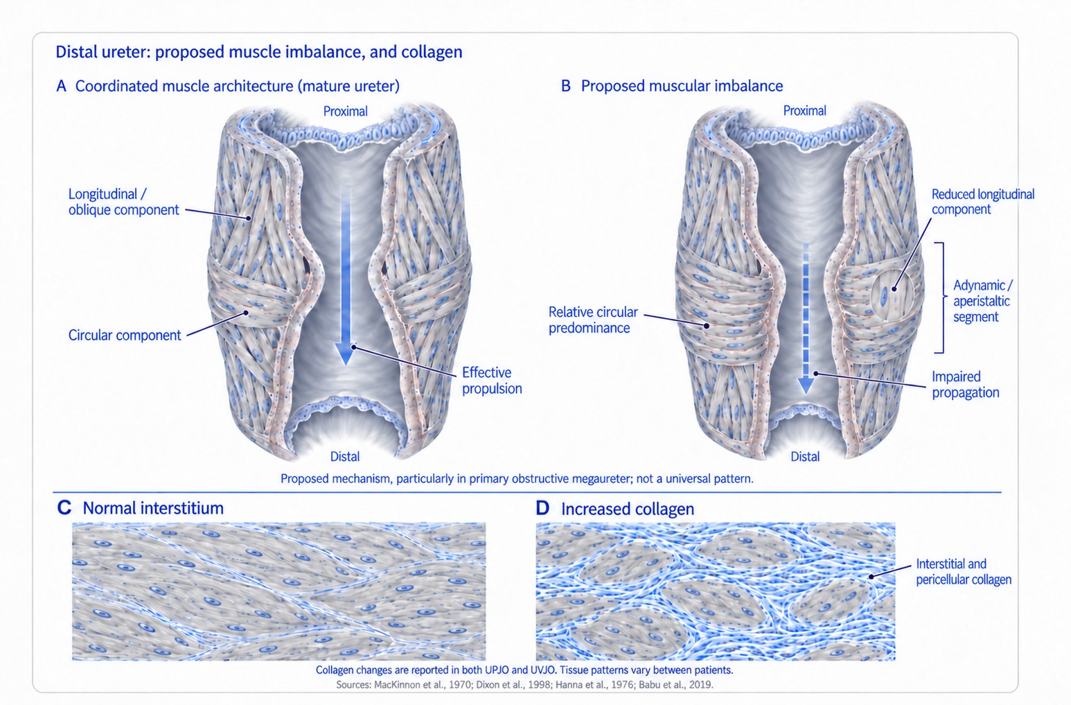
The muscle findings in infant UPJO are described differently. Central are poor development and continuity of circularly oriented fascicles and abnormal arrangement, and classically the normal arrangement of obliquely winding fascicles has also been described as replaced by longitudinally oriented fascicles or fibrous tissue (Murnaghan, 1958; Kim, 2024). In stereo-morphometric studies, the muscle of the normal UPJ changed from a circular arrangement in the newborn to an oblique meshwork in the adult, and subepithelial longitudinal muscle appeared at about 2 years of age; at the UPJ in congenital hydronephrosis, these age-related changes were indistinct or absent, and segmental muscle hypoplasia, abnormal fascicle arrangement and absence of longitudinal muscle were observed (Kaneto et al., 1988, 1991). Because the normal neonatal UPJ is also predominantly circular, the key is not the proportion of a particular orientation but whether the arrangement and connectivity required for that age and location have matured.
Scroll horizontally to see the full figure
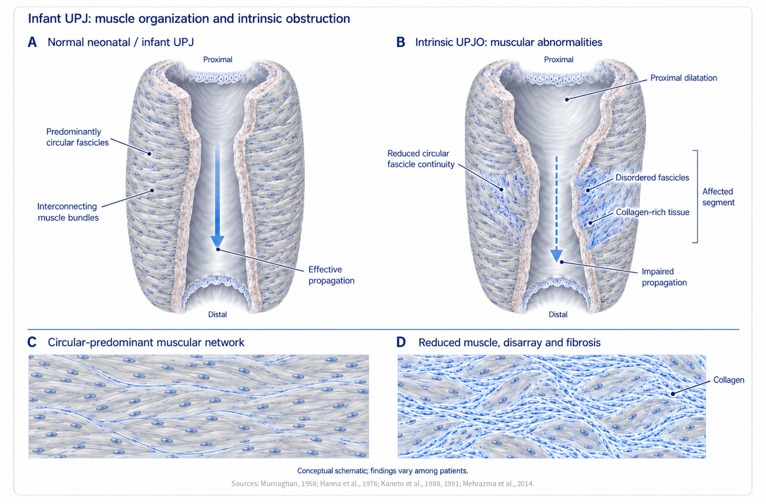
Increased collagen is a finding common to both sites. In an electron microscopy study, excess collagen between and around muscle cells and clusters of compromised muscle cells proximal to the collagenous segment were consistently observed in all 48 specimens of UPJO and primary obstructive megaureter, and the authors considered that this disrupts functional continuity through intercellular junctions (Hanna et al., 1976). A study comparing 25 UPJO cases with 19 autopsy controls likewise found more collagen deposition, irregular fascicle arrangement and fewer Cajal-like cells (Mehrazma et al., 2014). Abnormal muscle arrangement and increased collagen can occur together, but this does not prove a common cause.
Babu et al. (2019) compared normal ureters (20 cases), the dilated portion and the narrow segment of UVJO specimens (14 cases), the UPJO narrow segment excised at pyeloplasty (31 cases), and fetal ureters (12 cases). Compared with 0.75 in normal ureters, the collagen-to-muscle ratio was higher, at 3.0 in the UVJO narrow segment, 2.5 in the UPJO narrow segment and 3.1 in fetal ureters, and interstitial cells of Cajal were fewer, at 4.5, 5.1 and 5.0, respectively, compared with 16.1 per 10 high-power fields. The dilated ureter above the UVJO, by contrast, at 0.65 and 17.3, showed no statistically significant difference from normal (Figure 4). Another report, however, found increased collagen and elastic tissue throughout the dilated segment (Gosling and Dixon, 1978), so findings in the dilated portion vary between studies.
Scroll horizontally to see the full figure
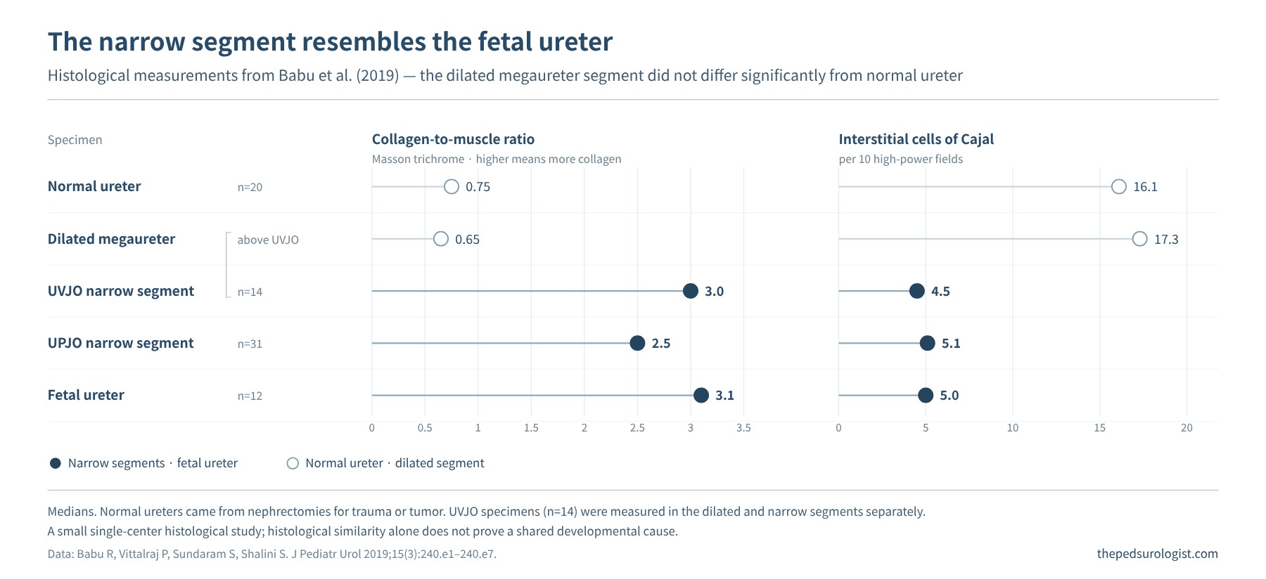
Based on the observation that in the fetal ureter interstitial cells of Cajal and smooth muscle increase and collagen decreases as gestation advances, the authors proposed a maturational arrest hypothesis, in which UPJO and UVJO may represent failure of this maturation to be completed at the two ends. This was, however, a small single-center histological comparison, and histological similarity alone does not prove a common developmental cause. In particular, fibrosis and muscle changes at the time of surgery may include both preexisting abnormalities and changes that followed impaired drainage.
In human fetal studies, ureteral muscle first differentiated in a spiral orientation in the upper ureter at about 12 weeks of gestation, whereas the longitudinal muscle of the intravesical ureter began to form fascicles only at about 17 weeks (Matsuno et al., 1984). In a porcine study, the musculature and innervation of the ureterovesical junction continued to mature after birth (Pirker et al., 2007). This is used as evidence to explain transient UVJ dysfunction and spontaneous improvement of some megaureters. Babu et al. (2019), by contrast, proposed that maturation may start in the mid-ureter and progress toward both ends. Because the study subjects and the measures used to assess maturation differ, these are hard to merge into one settled direction. Here we focus on the shared view that "the ureteral wall and its transport function continue to mature even after the form has been established."
The same tissue change is not the same disease
UPJO and primary obstructive megaureter cause impaired drainage at different locations. Even when common histological findings are reported, this does not mean that all developmental mechanisms and the clinical course of the two conditions are the same. Infant UPJO is mostly an intrinsic abnormality of the junction itself, but with increasing age, extrinsic causes such as compression or kinking related to a lower-pole crossing vessel account for a larger share. In 31 adults with UPJO, accessory lower-pole vessels were found in 52%, twice the rate in children (Lowe and Marshall, 1984).
| Feature | UPJO | Primary obstructive megaureter |
|---|---|---|
| Main site | The junction where the renal pelvis continues into the ureter | Distal ureter near the bladder · UVJ |
| Typical dilation | Pelvic and calyceal dilation; ureteral dilation not prominent | Ureteral dilation; may be accompanied by dilation of the upper collecting system |
| Range of mechanisms | Intrinsic abnormalities of tissue and propagation of contraction; extrinsic compression or kinking (lower-pole crossing vessel), etc. | Distal adynamic segment and abnormal tissue composition |
| Role of embryology | Explains the background against which the form and function of the junction are established | Explains the relationship between distal ureteral maturation and transport function |
| Limits of interpretation | Not every UPJO can be explained by a single developmental defect | The form of megaureter alone cannot establish obstruction |
The adynamic segment referred to in primary obstructive megaureter is a segment of the distal ureter in which peristalsis is absent or not efficiently propagated. It does not mean that the lumen is completely closed. Megaureter should be considered in categories: refluxing, obstructed, non-refluxing non-obstructed, and combined refluxing and obstructed. The table above compares typical patterns and is not a criterion for diagnosing the cause from imaging findings alone.
Distal maturation is one of the hypotheses that explain spontaneous improvement of some megaureters. It does not, however, lead to the conclusion that "because the ureter is immature, all will improve with waiting." The actual course must be judged by following changes in dilation, infection, symptoms, drainage and renal function together.
What changes in the developing kidney
Impaired drainage in the fetus or newborn is different from pressure simply being added to an already completed kidney. Depending on the timing, degree and duration of obstruction, it can affect renal differentiation and nephron formation, and the subsequent injury response also differs. In experiments ligating the ureter in fetal sheep, obstruction in the first half of gestation caused renal dysplasia, and obstruction in the second half caused hydronephrosis (Beck, 1971).
When we see a kidney with thin parenchyma or low function, then, two questions should be kept side by side. Was the renal tissue insufficiently formed from the outset, and has additional injury arisen from impaired drainage? Both processes can coexist. Renal dysplasia and multicystic dysplastic kidney (MCDK) should not be placed simply as the end stage of severe hydronephrosis (Beck, 1971; Mackie and Stephens, 1975).
Embryology helps make these questions concrete. A single ultrasound or surgical specimen, however, cannot reconstruct the entire developmental process in reverse. Confirming the current state of the kidney and its change over time is still necessary.
Summary — the causes of dilation, seen through development
Seen through embryology, the causes of antenatal and infant urinary tract dilation fall into three groups according to which step went wrong. The first is an abnormal connection. Where the ureteric bud arises from the mesonephric duct, and where the ureter reconnects to the bladder after the common nephric duct is removed, determine duplex kidney, ectopic ureter, ureterocele and reflux; the further the orifice lay from its normal position, the more dysplastic the corresponding renal moiety (Mackie and Stephens, 1975; Batourina et al., 2005; Uetani et al., 2009). The second is abnormal differentiation and maturation of the ureteral wall. Even with an open lumen, peristalsis is not transmitted unless smooth muscle and interstitial cells of Cajal form a connected arrangement. Infant UPJO is described in terms of reduced continuity and disarray of circular fascicles, primary obstructive megaureter in terms of muscular imbalance in the adynamic segment, and increased collagen with fewer interstitial cells of Cajal has been reported in the narrow segment at both sites (Murnaghan, 1958; MacKinnon et al., 1970; Hanna et al., 1976; Kaneto et al., 1991; Babu et al., 2019). The third is the timing of normal maturation. Ureteral muscle differentiates from top to bottom and the segment near the bladder keeps maturing after birth, which explains some transient dilation and the spontaneous improvement of some megaureters (Matsuno et al., 1984; Pirker et al., 2007).
Abnormal parenchymal development is not a cause of dilation but an accompanying result of the same developmental error or of early obstruction. Dysplasia was more severe in moieties whose orifice lay away from its normal position (Mackie and Stephens, 1975), and obstruction early in development can produce dysplasia before dilation (Beck, 1971). A kidney underformed from the start can therefore look on imaging like one damaged by impaired drainage, and the cause of dilation and the state of the parenchyma need to be assessed separately.
These three groups are not mutually exclusive, and much of the evidence comes from animal models or small tissue studies. Embryology makes us ask where and why dilation arose, but whether an individual child is obstructed, and how to treat it, is still decided by imaging and functional studies over time.
Next — Ureteropelvic Junction Obstruction: When to Observe and When to Operate. We examine how to read ultrasound changes and diuretic renography, and how to judge an obstruction that threatens renal function.
References
- Schuchardt A, D'Agati V, Larsson-Blomberg L, Costantini F, Pachnis V. Defects in the kidney and enteric nervous system of mice lacking the tyrosine kinase receptor Ret. Nature. 1994;367(6461):380-383. doi:10.1038/367380a0
- Pichel JG, Shen L, Sheng HZ, et al. Defects in enteric innervation and kidney development in mice lacking GDNF. Nature. 1996;382(6586):73-76. doi:10.1038/382073a0
- Airik R, Bussen M, Singh MK, Petry M, Kispert A. Tbx18 regulates the development of the ureteral mesenchyme. J Clin Invest. 2006;116(3):663-674. doi:10.1172/JCI26027
- Mackie GG, Stephens FD. Duplex kidneys: a correlation of renal dysplasia with position of the ureteral orifice. J Urol. 1975;114(2):274-280. doi:10.1016/s0022-5347(17)67007-1
- Batourina E, Tsai S, Lambert S, et al. Apoptosis induced by vitamin A signaling is crucial for connecting the ureters to the bladder. Nat Genet. 2005;37(10):1082-1089. doi:10.1038/ng1645
- Viana R, Batourina E, Huang H, et al. The development of the bladder trigone, the center of the anti-reflux mechanism. Development. 2007;134(20):3763-3769. doi:10.1242/dev.011270
- Uetani N, Bertozzi K, Chagnon MJ, Hendriks W, Tremblay ML, Bouchard M. Maturation of ureter-bladder connection in mice is controlled by LAR family receptor protein tyrosine phosphatases. J Clin Invest. 2009;119(4):924-935. doi:10.1172/JCI37196
- Babu R, Vittalraj P, Sundaram S, Shalini S. Pathological changes in ureterovesical and ureteropelvic junction obstruction explained by fetal ureter histology. J Pediatr Urol. 2019;15(3):240.e1-240.e7. doi:10.1016/j.jpurol.2019.02.001
- Murnaghan GF. The dynamics of the renal pelvis and ureter with reference to congenital hydronephrosis. Br J Urol. 1958;30(3):321-329. doi:10.1111/j.1464-410x.1958.tb03525.x
- MacKinnon KJ, Foote JW, Wiglesworth FW, Blennerhassett JB. The pathology of the adynamic distal ureteral segment. J Urol. 1970;103(2):134-137. doi:10.1016/s0022-5347(17)61908-6
- Kim SW. Management of hydronephrosis: a comprehensive review in pediatric urology perspective. Child Kidney Dis. 2024;28(2):59-65. doi:10.3339/ckd.24.011
- Dixon JS, Jen PY, Yeung CK, Gosling JA. The vesico-ureteric junction in three cases of primary obstructive megaureter associated with ectopic ureteric insertion. Br J Urol. 1998;81(4):580-584. doi:10.1046/j.1464-410x.1998.00610.x
- Kaneto H, Chiba T, Takahashi T, Orikasa S. 腎盂尿管移行部における正常尿管壁の筋層構築 [Muscular architecture of the normal ureteral wall at the ureteropelvic junction: a three-dimensional morphometric study]. Jpn J Urol. 1988;79(6):1040-1048. doi:10.5980/jpnjurol1928.79.6_1040
- Kaneto H, Orikasa S, Chiba T, Takahashi T. Three-D muscular arrangement at the ureteropelvic junction and its changes in congenital hydronephrosis: a stereo-morphometric study. J Urol. 1991;146(3):909-914. doi:10.1016/s0022-5347(17)37959-4
- Hanna MK, Jeffs RD, Sturgess JM, Barkin M. Ureteral structure and ultrastructure. Part II. Congenital ureteropelvic junction obstruction and primary obstructive megaureter. J Urol. 1976;116(6):725-730. doi:10.1016/s0022-5347(17)58987-9
- Mehrazma M, Tanzifi P, Rakhshani N. Changes in structure, interstitial Cajal-like cells and apoptosis of smooth muscle cells in congenital ureteropelvic junction obstruction. Iran J Pediatr. 2014;24(1):105-110. PMC4359592
- Gosling JA, Dixon JS. Functional obstruction of the ureter and renal pelvis: a histological and electron microscopic study. Br J Urol. 1978;50(3):145-152. doi:10.1111/j.1464-410x.1978.tb02790.x
- Matsuno T, Tokunaka S, Koyanagi T. Muscular development in the urinary tract. J Urol. 1984;132(1):148-152. doi:10.1016/s0022-5347(17)49508-5
- Pirker ME, Rolle U, Shinkai T, Shinkai M, Puri P. Prenatal and postnatal neuromuscular development of the ureterovesical junction. J Urol. 2007;177(4):1546-1551. doi:10.1016/j.juro.2006.11.081
- Lowe FC, Marshall FF. Ureteropelvic junction obstruction in adults. Urology. 1984;23(4):331-335. doi:10.1016/0090-4295(84)90133-x
- Beck AD. The effect of intra-uterine urinary obstruction upon the development of the fetal kidney. J Urol. 1971;105(6):784-789. doi:10.1016/s0022-5347(17)61629-x