
As Part 1 showed, a hydronephrosis grade tells you about likelihood, not cause. Postnatal evaluation therefore has three aims: to confirm that the dilation actually persists, to distinguish reflux from obstruction from a lower urinary tract problem, and to protect the kidney and the child from infection while we wait (Kim, 2024).
These aims come down to three decisions: when to obtain the first ultrasound, who should have a VCUG and diuretic renography, and who should receive antibiotic prophylaxis. This article reads three documents separately: the UTD consensus (2014), which created the language of risk groups; the American Academy of Pediatrics (AAP) clinical report (2025), which built on that framework with the registry data accumulated since; and the EAU/ESPU guidelines (2026). Their answers to the same questions differ slightly, so unless it is clear which document a recommendation comes from, timing and target populations are easily conflated. Even in hospitals that record Onen grade, ureteral and bladder findings and infection risk must be assessed separately.
When to obtain the first ultrasound
For about the first two days of life, fluid redistributes out of the intravascular space in the newborn and urine output falls. An ultrasound in this window can make the dilation look smaller than it is. The UTD consensus, the AAP report and the EAU/ESPU guidelines therefore all place the first postnatal ultrasound after 48 hours of life.
How long it can wait depends on the prenatal risk group. AAP 2025 advises that a child who was A2–3 at any point prenatally should have the first ultrasound during the birth hospitalization, ideally after 48 hours, so that the need for antibiotic prophylaxis and further testing can be settled early. It adds, however, that discharge need not be delayed for this study unless bladder outlet obstruction is suspected. A child whose A1 persisted beyond 32 weeks is imaged between 48 hours and 6 weeks of life. The 2014 UTD consensus had proposed after 48 hours and within 1 month for both A1 persisting beyond 32 weeks and A2–3. In effect, AAP 2025 brought the first study forward for A2–3 and widened the window for A1 to 6 weeks.
The exception is when waiting is not an option. Severe bilateral dilation, a dilated solitary kidney, oligohydramnios, or suspected urethral obstruction call for evaluation immediately after birth. In particular, when bilateral dilation in a boy is accompanied by bladder wall thickening or a dilated posterior urethra, posterior urethral valves must be considered first, and this lies outside the follow-up framework of this article. The AAP report likewise excludes severe bilateral dilation with suspected bladder outlet obstruction from its scope, and directs that specialist consultation, bladder decompression, imaging and antibiotic prophylaxis begin within 48 hours of birth.
One more point: a child who qualifies for postnatal evaluation is imaged once more even if the first ultrasound is normal. AAP 2025 calls for at least two postnatal ultrasounds before follow-up ends, with one more at 3–6 months if the first showed no dilation. The basis is a report that in nearly half of children with prenatal dilation and a normal first postnatal ultrasound, the next ultrasound was abnormal again. The 2014 consensus proposed the second ultrasound 1–6 months later for prenatal A1.
The cases in which follow-up can be omitted are narrow. When the prenatal grade was A1 and the dilation resolved on ultrasound after 32 weeks, both the 2014 UTD consensus and AAP 2025 considered postnatal evaluation unnecessary. This exception should not be extended to prenatal A2–3; when the prenatal classification is unclear or undocumented, postnatal evaluation proceeds as for A1.
Follow-up by risk group — 2014 and 2025
The 2014 UTD consensus proposed follow-up intervals and additional tests for each postnatal risk group, but left most items "at the discretion of the clinician." The only items that could be called recommendations were VCUG and antibiotic prophylaxis in the high-risk group (P3). This was because the evidence at the time was insufficient.
| UTD consensus 2014 | Follow-up ultrasound | VCUG | Antibiotic prophylaxis | Functional imaging (diuretic renography) |
|---|---|---|---|---|
| P1 (low risk) | In 1–6 months | Clinician discretion | Clinician discretion | — |
| P2 (intermediate risk) | In 1–3 months | Clinician discretion | Clinician discretion | Clinician discretion |
| P3 (high risk) | Within 1 month | Recommended | Recommended | Clinician discretion |
Eleven years later, the AAP report made both ends explicit on the same risk groups. In P1 it does not recommend VCUG, functional imaging or antibiotic prophylaxis; in P3 it recommends all three. P2, in between, is still left to individual risk and shared decision-making with the family. And independently of risk group, it recommends VCUG and antibiotic prophylaxis for a child whose ureter is dilated to 7 mm or more.
| AAP 2025 | Follow-up ultrasound | VCUG | Antibiotic prophylaxis | Functional imaging (diuretic renography) |
|---|---|---|---|---|
| P1 (low risk) | In 3–6 months | Not recommended | Not recommended | Not recommended (reassess if dilation worsens) |
| P2 (intermediate risk) | In 1–3 months | Based on individual risk | Based on individual risk | Based on individual risk |
| P3 (high risk) | 1 month after the first ultrasound | Recommended | Recommended | Recommended (usually at 6–12 weeks of age if P3 persists) |
| Ureter ≥7 mm | — | Recommended | Recommended | — |
What to remember when reading these tables is that the conditions defining P1 carry the premise that the parenchyma, ureter and bladder are normal. A kidney with thinned parenchyma should not be grouped on the "no further testing needed" side simply because only the renal pelvis is dilated. That kidney is P3. Beyond that, decisions take in the degree of dilation and the parenchymal findings, together with whether the ureter is dilated, whether the bladder is normal, and which child this is (sex, circumcision status, history of febrile UTI). The three sections below set out the evidence.
What "based on individual risk" means in P2
AAP 2025 does not set a uniform rule for VCUG, antibiotic prophylaxis or functional imaging in P2; it asks that each child's risk be weighed and the decision made with the family, because the evidence is not as firm as for P1 or P3. Three main things go on the scale.
- Risk of UTI — when deciding on VCUG and antibiotic prophylaxis. The infection risk factors listed by the AAP are female sex, uncircumcised males (in P2 and P3), distal ureteral dilation of 7 mm or more, vesicoureteral reflux, and suspected obstructive uropathy such as bilateral dilation. Coexisting urinary tract anomalies such as a duplex ureter, solitary kidney, multicystic dysplastic kidney or neurogenic bladder place the child toward the higher-risk side. Of these, a ureter of 7 mm or more is not a matter of individual judgment but a condition for which VCUG and antibiotic prophylaxis are recommended, and if a febrile UTI occurs even once, reflux is assessed with VCUG.
- Risk of obstruction and functional loss — when deciding on functional imaging. On follow-up, look at whether the dilation increases, whether peripheral calyceal dilation becomes more pronounced, and whether the parenchymal findings change. In hydronephrosis with a UPJ obstruction pattern, a postnatal APD of 15 mm has been reported as a predictor of the likelihood of surgery, but this value alone does not decide testing or surgery. With severe bilateral dilation, bilateral parenchymal abnormality or a solitary functioning kidney, overall renal function is assessed as well.
- The burden of testing and treatment, and the family's choice. VCUG requires catheterization and radiation, and antibiotic prophylaxis carries the burden of resistant organisms and adverse effects. The decision is made after explaining the likelihood of future UTI, the likelihood of finding reflux and how it would then be treated, and the natural history of reflux and dilation.
Scroll horizontally to see the full figure
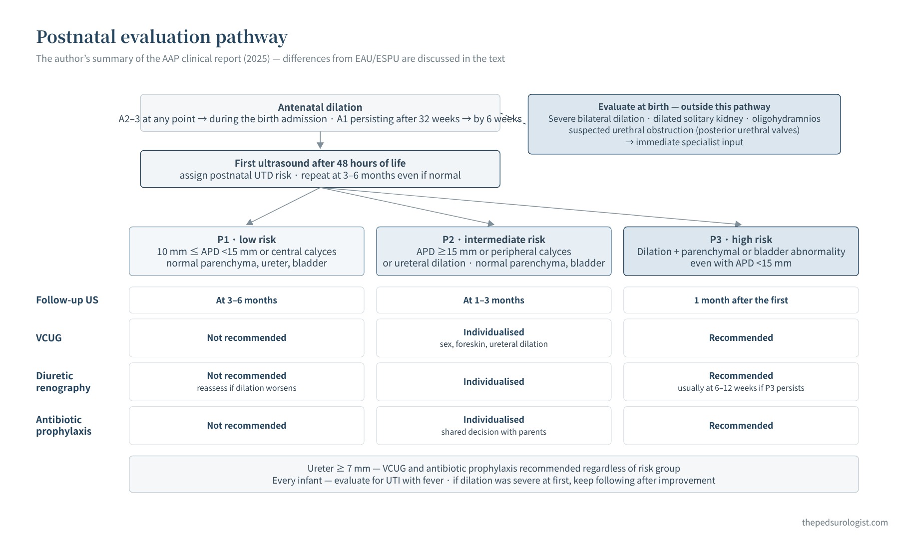
Who needs a VCUG
The dilemma around VCUG starts from the fact seen at the end of Part 1. The degree of prenatal dilation is proportional to the risk of a pathological cause after birth, but the risk of reflux did not differ appreciably with the degree of dilation (Lee et al., 2006). Grade, then, cannot select who needs a VCUG, and conversely there is no reason to perform one in every child, because detecting low-grade reflux in an asymptomatic infant seldom changes management (VUR guidelines, Part 1).
So the question shifts from "is there reflux?" to "is this a child in whom the reflux found would change management?" AAP 2025 likewise does not regard dilation itself as an indication for VCUG in asymptomatic neonates and infants, and makes the risk of UTI the main criterion for the VCUG decision. As in the table above, it is not recommended in P1 and is recommended in P3 and for a ureter of 7 mm or more; in P2 it is discussed with the family, weighing risk factors such as sex, circumcision status and ureteral dilation and the benefit of antibiotic prophylaxis. If a child with dilation develops a febrile UTI, reflux is assessed with VCUG.
The EAU/ESPU guidelines advise considering VCUG after a UTI, when the ureter is dilated, when there is a bladder abnormality, and in boys with bilateral dilation, and state that it is usually not needed in unilateral hydronephrosis without ureteral dilation or bladder abnormality. Routine VCUG before pyeloplasty also does not change surgical outcomes, so they advise performing it selectively.
The infection-risk data point the same way. In the prospective study by Braga et al. (2015) following 334 infants with prenatal hydronephrosis, febrile UTI occurred in 19% (median age 4 months), and the factors most strongly associated were reflux (HR 20.8) and hydronephrosis with ureteral dilation (HR 10.9). Hazard ratios from an observational study do not directly prove the benefit of a test, but identifying reflux in a child with a dilated ureter changes the infection-prevention plan, whereas in a child with pelvic dilation alone it often does not.
Diuretic renography: when and how
Diuretic renography (MAG3) assesses differential renal function and drainage. In the first few months of life the kidney is immature, tracer uptake is low and cortical transit is slow, so the results are unreliable. The study is therefore deferred: EAU/ESPU 2026 gives after 4–6 weeks of age, and AAP 2025 no earlier than 6–12 weeks of age. Indications are parenchymal abnormality, persistent or worsening severe dilation, and suspected obstruction; it is not performed in P1 merely because dilation persists. The 2014 consensus had left functional imaging in P2 and P3 to clinician discretion.
Diuretic renography results vary considerably with the conditions of the study. Hydration status, bladder filling, the timing of diuretic administration and the way regions of interest are drawn all affect differential function and the drainage curve. In practice, follow the pediatric standard protocol of your nuclear medicine department, but keep in mind that comparing two studies of the same child acquired under different conditions can lead to misreading a change in function.
Adequate hydration before the study; intravenous normal saline 15 mL/kg over 30 minutes, starting 15 minutes before radiopharmaceutical injection, then maintenance at 4 mL/kg per hour; bladder catheterization; and furosemide (Lasix) 1 mg/kg in the first year of life and 0.5 mg/kg at 1–16 years (maximum 40 mg). AAP 2025 describes giving 1 mg/kg (maximum 40 mg) if tracer remains after 15–20 minutes. Whether to catheterize every child varies between nuclear medicine procedure guidelines and between institutions, so follow your institution's standard procedure for protocol details.
How to interpret the results and when to proceed to surgery is covered in the article on UPJ obstruction. The topics there are how to read differential function below 40%, a decline of more than 10 percentage points on follow-up, the drainage curve, and a differential function measured as high as 55% or more, together with other findings. An example of a decline of more than 10 percentage points is a fall from 45% to 34%, which is not the same as a reduction by 10% of 45%.
Antibiotic prophylaxis — how far does the evidence go
There was a time when antibiotic prophylaxis was given routinely to children with prenatal hydronephrosis. This was less a matter of evidence than a practice carried over from children with reflux. That practice has now largely been abandoned, but who should receive prophylaxis is still debated. Randomized trials exist, but they are small and their populations are limited. The placebo-controlled trial by Braga et al. (2014) in infants with high-grade hydronephrosis was a pilot study that enrolled 46 infants and stopped at establishing whether a definitive trial was feasible. In the trial by Rianthavorn and Phithaklimnuwong (2020), which randomized 80 infants with mild to moderate isolated hydronephrosis, UTI occurred in 14.7% of the antibiotic group and 10.0% of the observation group, showing no benefit; among the UTIs that occurred, the proportion caused by cotrimoxazole-resistant organisms was 4 times higher in the antibiotic group (RR 4.0), and the trial was stopped early. Neither result can be applied as is to high-risk children with a dilated ureter.
Most decisions therefore rest on systematic reviews and observational cohorts. Children who did and did not receive antibiotics may have differed in risk from the outset, so the numbers must be read with patient selection and possible confounding in mind.
Scroll horizontally to see the full figure
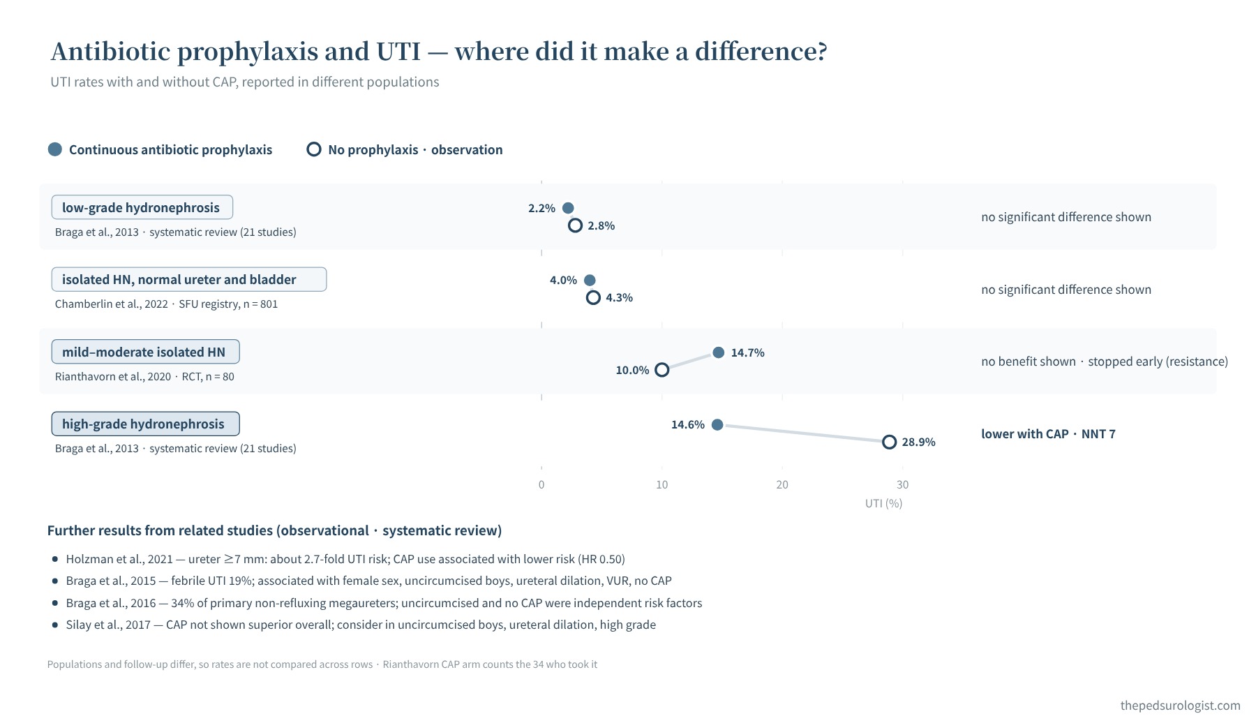
Divided by population, the evidence in the figure is as follows.
- Studies of low-grade and isolated hydronephrosis — no significant preventive effect shown. Low-grade hydronephrosis: 2.2% vs 2.8% (Braga et al., 2013, systematic review of 21 studies, 3,876 infants); isolated hydronephrosis without ureteral or bladder abnormality in 801 children: 4.0% vs 4.3% (Chamberlin et al., 2022). The latter included 35% with SFU grade 3–4, and no significant reduction was shown in the female, uncircumcised male or high-grade subgroups either. This does not apply to P3 with parenchymal or bladder abnormality.
- High-risk groups — a difference. High-grade hydronephrosis: 14.6% vs 28.9%, with a number needed to treat of 7 to prevent one infection (Braga et al., 2013, no randomized trials among the included studies). A ureter of 7 mm or more carried about 2.7 times the risk of UTI, and antibiotic use was associated with lower risk (Holzman et al., 2021, HR 0.50).
- Factors that raised risk. Female sex, uncircumcised males, ureteral dilation, reflux, and no antibiotic prophylaxis (Braga et al., 2015, febrile UTI in 19% of 334). In primary non-refluxing megaureter, febrile UTI occurred in 34%, mostly within the first 6 months of life (Braga et al., 2016).
- Conclusion of the systematic review. Overall, antibiotics cannot be said to be better than observation, but they are worth considering in uncircumcised boys, ureteral dilation and high-grade hydronephrosis (Silay et al., 2017, EAU/ESPU panel).
In short, isolated hydronephrosis without ureteral or bladder abnormality carries a low baseline risk of infection (about 4%), and no benefit of antibiotics has been shown. This does not extend to P3 with parenchymal abnormality. Failure to show a difference is not proof of no benefit, but there is no basis for routine prophylaxis in this group. Conversely, ureteral dilation (particularly 7 mm or more) or reflux carries a higher infection risk, and antibiotic use has been reported together with lower infection rates (Herz et al., 2014; Holzman et al., 2021). Sex and the foreskin raise risk, but because the AAP lists the foreskin as a risk factor in P2 and P3, these alone do not convert P1 to high risk.
The recommendations differ in population and strength between documents. EAU/ESPU 2026 does not recommend antibiotic prophylaxis for asymptomatic UPJ obstruction and suggests it for high-risk groups such as uncircumcised infants, hydronephrosis with ureteral dilation and high-grade hydronephrosis (weak recommendation). AAP 2025 recommends it in P3 and for a ureter of 7 mm or more, leaves P2 to individual risk and the family's choice, and does not recommend it in P1. The 2014 UTD consensus recommended it in P3 and left the rest to discretion. It is important not to merge these differences into a single sentence.
The burden of antibiotics also belongs on the scale. Long-term use raises concerns about increasing resistant organisms, adverse effects and changes in the gut microbiome, and whether antibiotics reduce renal scarring in prenatal hydronephrosis has not yet been studied. The real question is therefore not "whether to give it" but "to whom, and for how long." Most febrile UTIs (89% in the SFU registry; Zee et al., 2016) occurred within the first year of life, and the AAP advises considering discontinuation at 12 months of age unless reflux or ureteral dilation of 7 mm or more persists. It is better to set the timing of reassessment and the conditions for stopping when prophylaxis is started.
Circumcision and foreskin care
In boys, the foreskin comes up repeatedly as a risk factor for UTI. In the study by Ellison et al. (2018), which analyzed 5,561 boys with a hydronephrosis-related diagnosis in US insurance claims data, the odds of UTI in the first year were lower in the neonatal circumcision group (OR 0.36). The number needed to treat calculated by the authors was 10 with hydronephrosis and 83 in healthy children. These are odds ratios from an observational study, however, and the number needed to treat depends on the baseline risk of the population. The AAP report also mentions studies in which circumcision showed a protective effect similar to that of antibiotic prophylaxis, and the EAU/ESPU guidelines cite a systematic review showing that circumcision reduces UTI in boys with isolated prenatal hydronephrosis.
In Korea, however, neonatal circumcision is uncommon, and circumcision is irreversible. Rather than carrying this over as grounds for recommending it to every boy with hydronephrosis, discuss it with the family as one option in high-risk boys with ureteral dilation or reflux. The basic rule is not to forcibly retract the foreskin, and topical steroids are decided on after the condition of the foreskin and the indication have been confirmed in clinic. A parent-facing explanation of the foreskin is in "His foreskin balloons when he pees".
Summary — the ureter, the parenchyma and the child, rather than the size of the dilation
In postnatal evaluation, what decides management is less how many millimeters the renal pelvis measures than whether the ureter is dilated, whether the parenchyma and bladder are normal, and which child this is. Infection risk is judged from the ureteral and bladder findings together with individual risk factors; the risk of obstruction and functional loss from the degree and change of dilation together with the parenchymal findings. Low-risk dilation with normal parenchyma, ureter and bladder is managed mainly with ultrasound follow-up, and VCUG and antibiotic prophylaxis are usually unnecessary. When the ureteral diameter is 7 mm or more, or the child is P3, VCUG and antibiotic prophylaxis are recommended. Functional imaging is recommended in P3, and in P2 it is decided individually according to the degree of suspected obstruction and the follow-up findings. Both the initial risk group and changes on follow-up enter the decision.
The first postnatal ultrasound is obtained after 48 hours of life. AAP 2025 proposes, by prenatal risk group, the birth hospitalization (A2–3) or within 6 weeks of life (persistent A1); the 2014 UTD consensus proposed within 1 month. P1 is observed, P2 individualized, and P3 and a ureter of 7 mm or more are evaluated actively. Stating which guideline a recommendation comes from reduces confusion.
Next — How the Ureter Forms, and Why It Dilates. Starting from the development of the ureteric bud, the connection between ureter and bladder, and the differentiation and maturation of the ureteral wall, we examine the mechanisms of UPJ obstruction and UVJ obstruction (primary obstructive megaureter). UPJ obstruction and megaureter then each get an article of their own.
References
- Nguyen HT, Benson CB, Bromley B, et al. Multidisciplinary consensus on the classification of prenatal and postnatal urinary tract dilation (UTD classification system). J Pediatr Urol. 2014;10(6):982-998. doi:10.1016/j.jpurol.2014.10.002
- EAU Guidelines on Paediatric Urology. Dilatation of the upper urinary tract (PUJ obstruction); Foetal urology. European Association of Urology, 2026.
- Herndon CDA, Otero HJ, Hains D, et al. Perinatal urinary tract dilation: recommendations on pre-/postnatal imaging, prophylactic antibiotics, and follow-up: clinical report. Pediatrics. 2025;156(1):e2025071814. doi:10.1542/peds.2025-071814
- Lee RS, Cendron M, Kinnamon DD, Nguyen HT. Antenatal hydronephrosis as a predictor of postnatal outcome: a meta-analysis. Pediatrics. 2006;118(2):586-593. doi:10.1542/peds.2006-0120
- Braga LH, Farrokhyar F, D'Cruz J, Pemberton J, Lorenzo AJ. Risk factors for febrile urinary tract infection in children with prenatal hydronephrosis: a prospective study. J Urol. 2015;193(5 Suppl):1766-1771. doi:10.1016/j.juro.2014.10.091
- Braga LH, Pemberton J, Heaman J, DeMaria J, Lorenzo AJ. Pilot randomized, placebo controlled trial to investigate the effect of antibiotic prophylaxis on the rate of urinary tract infection in infants with prenatal hydronephrosis. J Urol. 2014;191(5 Suppl):1501-1507. doi:10.1016/j.juro.2013.10.033
- Braga LH, Mijovic H, Farrokhyar F, Pemberton J, DeMaria J, Lorenzo AJ. Antibiotic prophylaxis for urinary tract infections in antenatal hydronephrosis. Pediatrics. 2013;131(1):e251-e261. doi:10.1542/peds.2012-1870
- Rianthavorn P, Phithaklimnuwong S. The role of antibiotic prophylaxis in mild to moderate isolated hydronephrosis detected in antenatal screening. Investig Clin Urol. 2020;61(2):200-206. doi:10.4111/icu.2020.61.2.200
- Silay MS, Undre S, Nambiar AK, et al. Role of antibiotic prophylaxis in antenatal hydronephrosis: a systematic review from the European Association of Urology/European Society for Paediatric Urology Guidelines Panel. J Pediatr Urol. 2017;13(3):306-315. doi:10.1016/j.jpurol.2017.02.023
- Chamberlin JD, Braga LH, Davis-Dao CA, et al. Continuous antibiotic prophylaxis in isolated prenatal hydronephrosis. J Pediatr Urol. 2022;18(3):363.e1-363.e7. doi:10.1016/j.jpurol.2022.03.027
- Holzman SA, Braga LH, Zee RS, et al. Risk of urinary tract infection in patients with hydroureter: an analysis from the Society of Fetal Urology Prenatal Hydronephrosis Registry. J Pediatr Urol. 2021;17(6):775-781. doi:10.1016/j.jpurol.2021.09.001
- Zee RS, Herbst KW, Kim C, et al. Urinary tract infections in children with prenatal hydronephrosis: a risk assessment from the Society for Fetal Urology Hydronephrosis Registry. J Pediatr Urol. 2016;12(4):261.e1-261.e7. doi:10.1016/j.jpurol.2016.04.024
- Herz D, Merguerian P, McQuiston L. Continuous antibiotic prophylaxis reduces the risk of febrile UTI in children with asymptomatic antenatal hydronephrosis with either ureteral dilation, high-grade vesicoureteral reflux, or ureterovesical junction obstruction. J Pediatr Urol. 2014;10(4):650-654. doi:10.1016/j.jpurol.2014.06.009
- Braga LH, D'Cruz J, Rickard M, Jegatheeswaran K, Lorenzo AJ. The fate of primary nonrefluxing megaureter: a prospective outcome analysis of the rate of urinary tract infections, surgical indications and time to resolution. J Urol. 2016;195(4 Pt 2):1300-1305. doi:10.1016/j.juro.2015.11.049
- Ellison JS, Dy GW, Fu BC, Holt SK, Gore JL, Merguerian PA. Neonatal circumcision and urinary tract infections in infants with hydronephrosis. Pediatrics. 2018;142(1):e20173703. doi:10.1542/peds.2017-3703
- Kim SW. Management of hydronephrosis: a comprehensive review in pediatric urology perspective. Child Kidney Dis. 2024;28(2):59-65. doi:10.3339/ckd.24.011