
What we see before an operation is the result: where the meatus opened, how much the penis curves, how the skin is distributed. Why those three almost always travel together is only explained by looking at how the penis was built.
This instalment covers how the urethra forms — including the fact that shaft and glans are built in different ways — and what is left behind when that process stops partway. Prevalence and aetiology are taken up in Part 5.
The shaft urethra is built in two steps
In the penile shaft, two steps happen in order. The plate canalizes and a groove opens on the surface; the edges of that groove then meet and fuse into a tube. The glanular urethra is built by a different mechanism, taken up in the next section. First, how the two-step model came to be accepted.
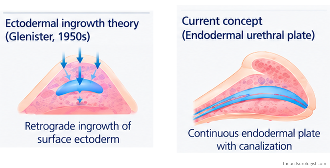
The account that held for decades was Glenister's ectodermal intrusion theory of 1954: surface ectoderm grows inward from the glans and meets the endodermal urethral plate advancing from the shaft, the two joining at the corona. The model had real explanatory appeal — it accounted for why hypospadias is commonest at the corona of all places. That is exactly where the two tissues were supposed to meet.
Recent work does not support it. Human fetal studies by Baskin, Cunha and colleagues have been put forward in support of the entire urethra deriving from the endodermal urethral plate, backed by immunostaining for the endodermal marker FOXA1. No evidence of ectoderm growing in retrograde was found.
There is a second reversal inside that story. Around 2000, fusion itself came under suspicion. Pig fetal studies failed to produce morphological evidence of midline fusion, and studies looking for epithelial-mesenchymal transformation — a marker of fusion — found none in urethral formation. The perineal raphe is often cited as evidence of fusion, but it was pointed out that the raphe alone does not settle the question.
As scanning electron microscopy and serial sectioning of human fetal tissue accumulated, fusion was reinstated, and what emerged was the double zipper hypothesis.
Opening zipper — the solid urethral plate canalizes from within, progressing distally, and produces an open groove on the surface: the urethral groove. It runs like a zipper being pulled open. This step is androgen-independent and happens identically in female urethral development.
Closing zipper — the edges of that groove, the urethral folds, meet and fuse at the midline to form a tubular urethra. This step is androgen-dependent and occurs only in males. It is not a simple apposition but an interdigitation proceeding at different rates in different layers. Where the edges meet an epithelial seam forms, then remodels away, leaving only the raphe at the surface.
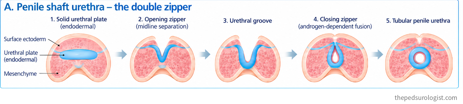
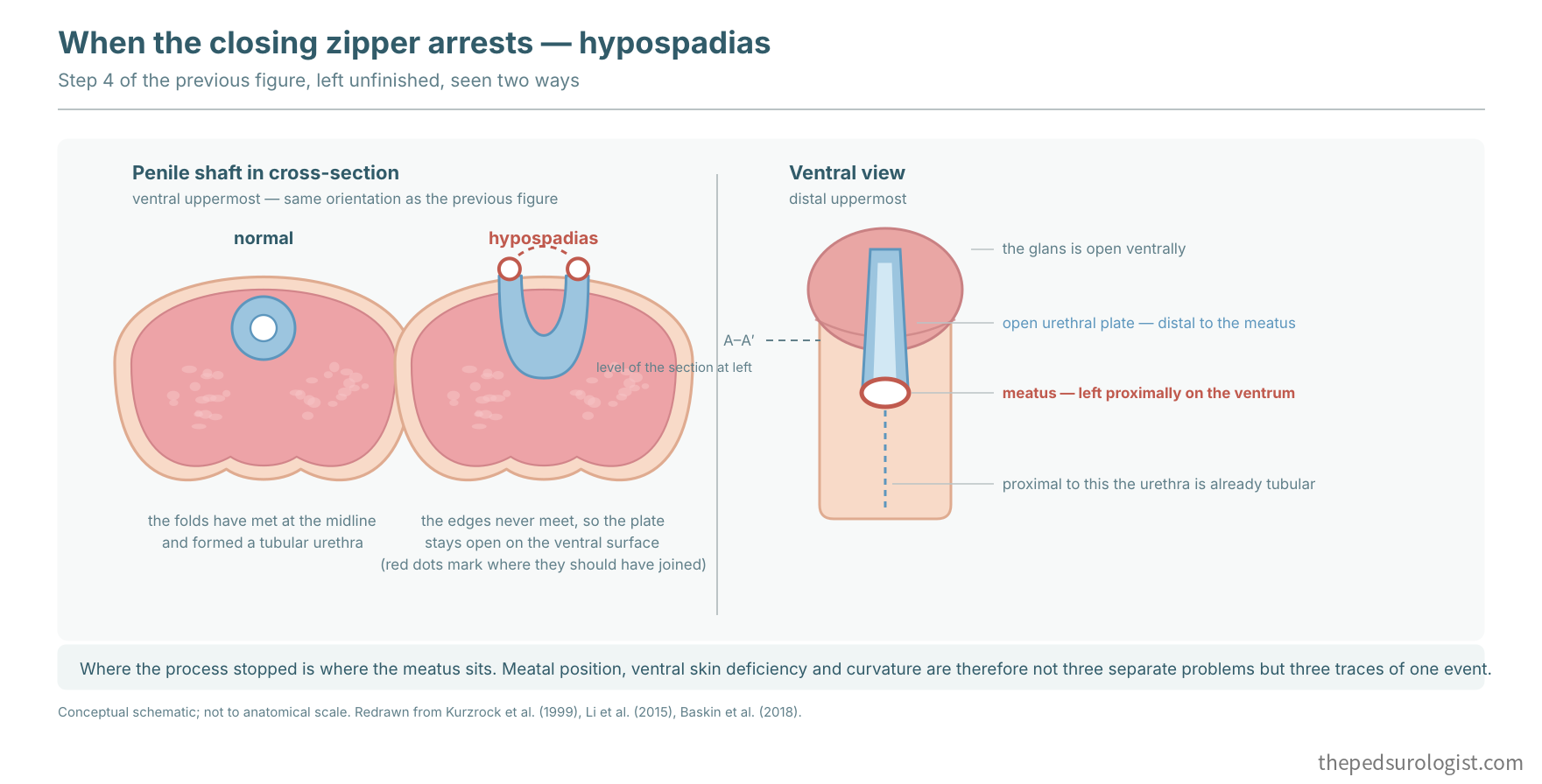
The order matters and is easy to read backwards. The urethra ends up beneath the skin because of fusion, not before it. At steps 2 and 3 the groove is plainly open to the exterior; at step 4 that open surface is covered, and only then does the urethra become an internal structure. The line where it was covered is the epithelial seam, and what remains after the seam remodels away is the raphe.
So canalization and fusion are two different processes, and androgen acts on the second. Where the androgen signal falls short, the urethra opens but never closes. The reason hypospadias leaves the ventrum open is contained in that sequence.
Why the two steps divide the way they do is clearer alongside female external genital development. Both sexes start from the same three structures — the genital tubercle, the urethral folds and the labioscrotal swellings. Without androgen the urethral folds never fuse and become the labia minora, the labioscrotal swellings never fuse and become the labia majora, and the midline stays open. One of the key events separating male from female external genital differentiation is whether the urethral folds and the labioscrotal swellings fuse at the midline.
Hypospadias can therefore be read as one phenotype in which ventral masculinization and fusion did not complete. The bifid scrotum seen in severe proximal cases follows the same account — the labioscrotal swellings failed to meet at the midline. The urethral folds and the labioscrotal swellings are neighbouring structures reading the same androgen signal, and when the signal is short, both are affected.
The glans is built by a different mechanism
What follows matters more in practice. The shaft urethra and the glanular urethra do not form the same way.
In the shaft the two steps above happen in sequence. Within the glans, however, the urethral plate canalizes in place without ever passing through an open groove, producing an independent tubular urethra. According to human fetal studies, there is neither an opening nor a closing zipper within the glans. Some observers have described a transient groove in the glans as well, however, so how the glanular urethra is completed is not yet fully agreed. Across weeks 12 to 18, canalization is followed by mesenchymal confluence over it and by remodeling of the surface epithelium.
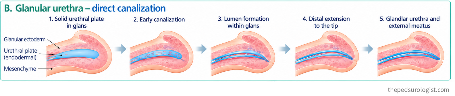
Baskin and colleagues set out five processes that must complete in order for the glanular urethra to form properly: the urethral plate must reach the tip of the glans and contact the surface epithelium; it must canalize; the lumen must extend to the tip; and mesenchymal confluence and epithelial remodeling must then complete.
This difference also changes how the position of the meatus should be read. Hypospadias of the shaft is explained fairly naturally as the point where the closing zipper stopped, but the same explanation is hard to extend to the glans, where there is no open groove to close in the first place. Yet in distal hypospadias an open glanular groove remains distal to the meatus. That suggests the failure in the glans lies less in canalization itself than in incomplete remodeling of the ventral epithelial seam and mesenchymal confluence.
The coronal region is more interesting still. Liu and colleagues pointed out that the commonest site of hypospadias in humans lies at the boundary where the two mechanisms meet. The very observation that Glenister's model explained as the place where two tissues meet, the current model explains again as the place where two mechanisms join.
This is, however, an interpretation drawn from normal fetal development; no study has yet shown directly which step fails in hypospadiac tissue. Upstream regulation — androgen signalling and epithelial–mesenchymal interaction — acts on both regions. In terms of morphogenesis, glanular hypospadias stops at a different step from shaft hypospadias, but on current evidence it is better seen as the distal end of the same spectrum than as a separate condition.
The urethra inside the glans is, from the very beginning, a structure built differently from the one in the shaft. What we do when we reconstruct a glans — tubularize the plate and close glans wings over it — does not recapitulate that process; it reaches a similar result by another route. A suture line is created, and nothing substitutes for mesenchyme converging over the tube. This is probably not unrelated to the way the G of the GMS score varies between raters, which Part 3 described. We judge the quality of glans and plate by eye, but we are not seeing at which developmental step that tissue stopped.
One corollary: urethral formation in the mouse differs from the human in important ways. Much of the mouse penile urethra forms by canalization of the plate in place — the same mechanism that operates in the human glans — so the opening and closing that take place in the human shaft are largely absent. Findings from mouse models therefore need care when applied to humans, particularly those concerning distal urethral morphogenesis. The point returns in the discussion of endocrine disruptors in Part 5.
Why the prepuce goes wrong with it
Part 1 argued that the abnormal meatal position, the curvature and the skin distribution are three traces of a single developmental event. The grounds are here.
The prepuce appears around week 8 as preputial folds at the base of the glans. It fuses dorsally first, and as the penis grows the folds rotate ventrally from both sides to enclose the glans. But the ventral surface closes last, and only after the urethral folds have fused. That is around week 20.
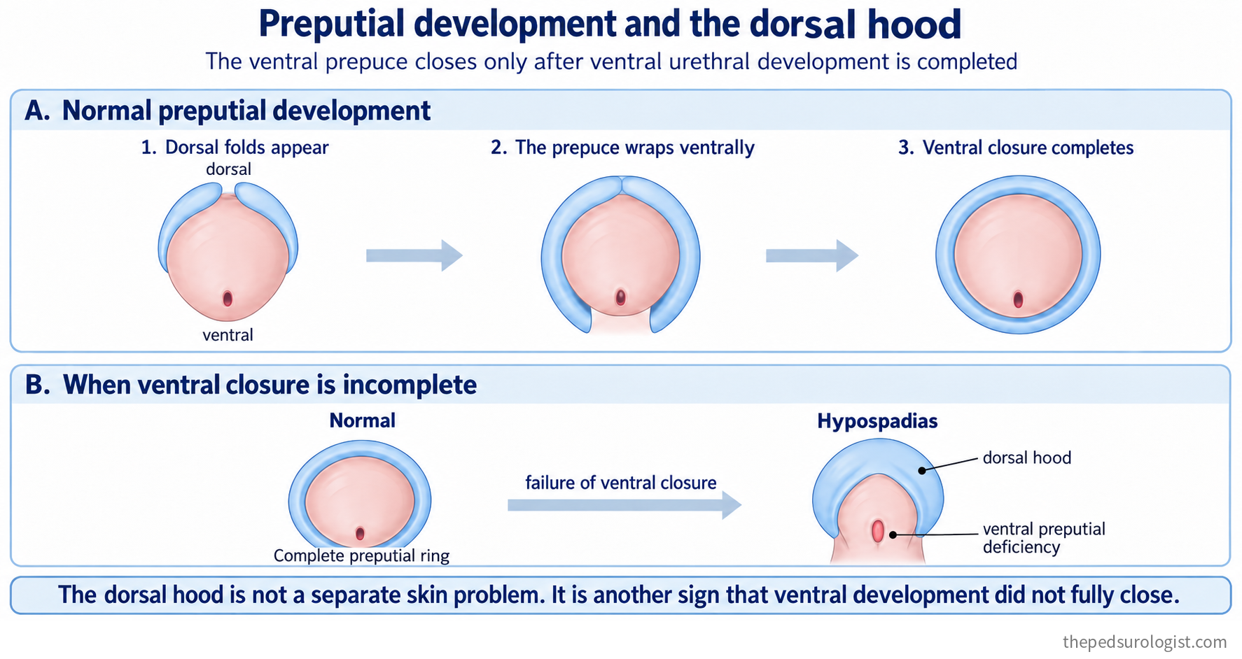
So if the urethral folds do not fuse all the way, neither does the prepuce ventrally. That is why a dorsal hood accompanies 95% of hypospadias cases. The dorsal hood is not an excess of skin dorsally; it is skin that never arrived ventrally.
The dorsal hood, in other words, is not an incidental finding alongside hypospadias but another expression of the same arrested process. The reason not to squander the prepuce at the primary repair is confirmed here as well — it is not spare skin but the residue of a process that never finished, and it is the only local tissue available for ventral reconstruction.
In the next instalment
Having seen how the urethra is built, the next instalment looks at how often that process goes wrong, and why.
Neither question has a simple answer. Prevalence reported by the 27 programmes of an international surveillance network ranges from 2.1 to 39.1 per 10,000 births — a nineteen-fold spread — and in a German study that actually examined meatal position in 500 adult men, 13% met the study definition of hypospadias. On the aetiological side, the most consistent association comes not from endocrine disruptors but from placental function. Part 5 takes it up.
References
- Glenister TW. The origin and fate of the urethral plate in man. J Anat. 1954;88(3):413-425.
- Kurzrock EA, Baskin LS, Cunha GR. Ontogeny of the male urethra: theory of endodermal differentiation. Differentiation. 1999;64(2):115-122.
- Baskin LS. Hypospadias and urethral development. J Urol. 2000;163(3):951-956.
- Hadidi AT, Roessler J, Coerdt W. Development of the human male urethra: a histochemical study on human embryos. J Pediatr Surg. 2014;49(7):1146-1152.
- Li Y, Sinclair A, Cao M, et al. Canalization of the urethral plate precedes fusion of the urethral folds during male penile urethral development: the double zipper hypothesis. J Urol. 2015;193(4):1353-1359.
- Baskin L, Shen J, Sinclair A, et al. Development of the human penis and clitoris. Differentiation. 2018;103:74-85.
- Liu G, Liu X, Shen J, Sinclair A, Baskin L, Cunha GR. Contrasting mechanisms of penile urethral formation in mouse and human. Differentiation. 2018;101:46-64.
- Penington EC, Hutson JM. Embryology of hypospadias. In: Hadidi AT, Azmy AF, eds. Hypospadias Surgery: An Illustrated Guide. Berlin: Springer; 2004.
- Hypospadias. In: Partin AW, Dmochowski RR, Kavoussi LR, Peters CA, eds. Campbell-Walsh-Wein Urology. 13th ed. Philadelphia: Elsevier. (Chapter 56)