
Parents ask about the cause less often than one might expect. When they do, it usually comes near the end of the visit, almost in passing — why did this happen, was there something they did during the pregnancy.
This instalment sets out how much of that question can be answered today: whether hypospadias is genuinely increasing, and how strong the evidence is behind the things that have been named as causes. The two steps of urethral formation described in Part 4 — canalization, then fusion — are the groundwork for what follows.
Is it really increasing?
The statement that hypospadias is on the rise is widespread. The evidence is less simple than that.
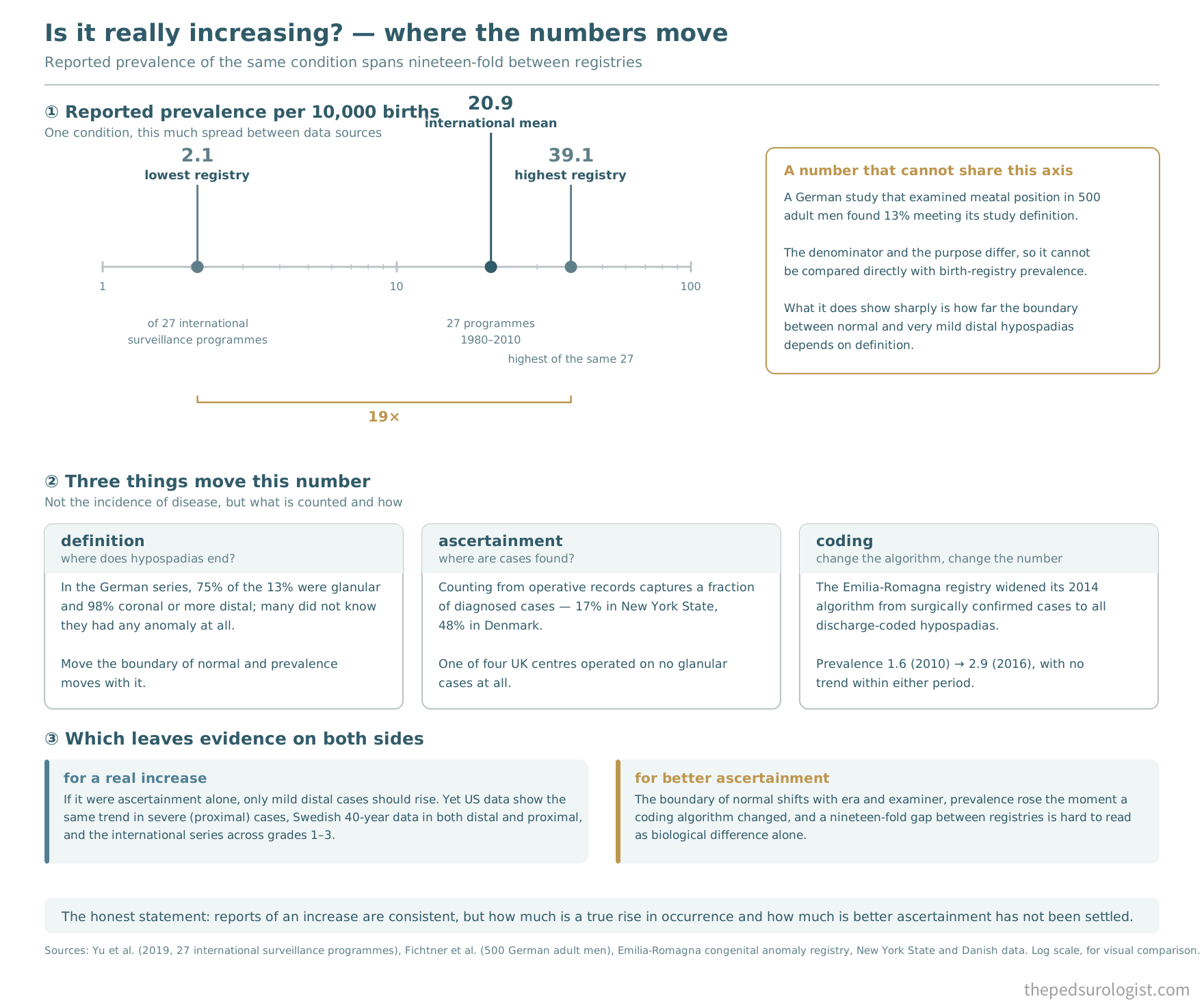
The largest dataset analysed 31 years, 1980 to 2010, across 27 programmes of an international birth defect surveillance network (Yu et al., 2019). Overall prevalence was 20.9 per 10,000 births, rising 1.6-fold over the study period (0.25 per 10,000 per year, p<0.05). A significant upward trend was seen in 61.9% of programmes, and the increase was present in grades 1, 2 and 3 alike. The authors noted this ran counter to earlier reports that the rise had levelled off.
In the same study, prevalence by programme ranged from 2.1 to 39.1 per 10,000. A nineteen-fold difference. It is hard to read all of that nineteen-fold gap as real biological difference. Much of it comes from what gets counted as hypospadias and how cases are found — whether mild distal cases are included, whether only surgically confirmed cases are counted, whether detection is at the newborn examination or from discharge coding.
One dataset shows where the boundary lies more starkly than any other. A German study that actually examined meatal position in 500 adult men found that 13% met its study definition of hypospadias. Of those, 75% were glanular and 98% coronal or more distal, and a substantial number, including men classified as coronal, did not know they had any penile anomaly at all.
That figure cannot be compared directly with birth-registry prevalence; the denominator and the purpose of the study are both different. What it does show sharply is how far the boundary between normal and very mild distal hypospadias depends on definition.
So are those men hypospadias patients? By the study definition yes; clinically no. It means there is no agreement on where normal ends. And if that boundary moves with the era and with the examiner, then some part of any change in prevalence is not disease becoming commoner but the range of normal becoming narrower. The Rochester series of 1940–1970, which reported a high prevalence of 4 per 1,000 while 87% of its cases were glanular or coronal, points the same way.
Systems that capture cases surgically introduce another variable. In New York State the operative rate was 0.6 per 1,000, implying that only about 17% of diagnosed cases were operated on; in Denmark the figure was 48%. In a UK survey, three of four centres had glanular cases making up a quarter of their operative volume, while one centre operated on no glanular cases at all. Mean age at surgery across European regions ranged from 13.6 to 60.3 months. In a system that counts patients from operative records, every one of those differences appears as a difference in prevalence.
One dataset tested this suspicion directly. In 2014 the Emilia-Romagna registry in Italy changed its algorithm, widening it from surgically confirmed cases to all hypospadias identified from discharge records. Prevalence rose from 1.6 in 2010 to 2.9 in 2016. Yet there was no significant year-to-year difference within 2010–2013, nor within 2014–2016; the only significant difference was between the two periods. The increase was the algorithm change.
There is evidence in the other direction too. If the rise were an artefact of ascertainment, only mild distal cases should be increasing. Yet US data show the same trend in severe (proximal) cases, 40 years of Swedish data report increases in both distal and proximal forms, and the international dataset cited above found increases across grades 1, 2 and 3. Some studies have reported annual increases of up to 3%.
So the honest statement is this. Reports of an increase are consistent, but how much of it is a true rise in occurrence and how much is better ascertainment has not been settled. That severe cases are also increasing argues for something real; that the boundary of normal moves and that coding methods change argue the other way. Both have evidence, and there is no conclusion yet.
What has been named as a cause
It helps to map the terrain first. In most cases no single cause can be identified. The placental-fetal environment, genetic susceptibility, environmental exposure and maternal factors act together, and cases explained by any one of them alone are a minority. The point of the model below is that these four domains converge on one place — the androgen pathway — and from there on failure of ventral urethral closure.
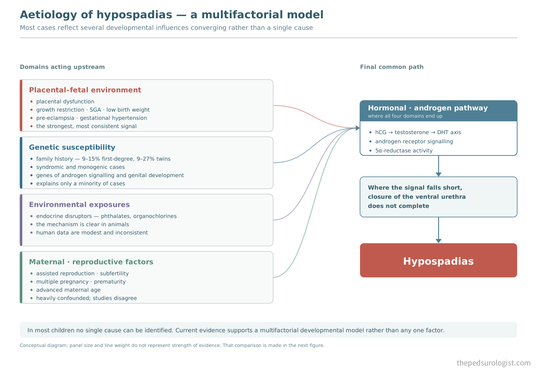
The four domains do not carry equal weight, however. The strength of evidence differs considerably between them. Taking the strongest first — but first, one hypothesis that ties them together.
The testicular dysgenesis hypothesis
Alongside reports that hypospadias is increasing came reports that cryptorchidism is increasing, semen quality is falling and testicular cancer is rising. In 2001 Skakkebaek and colleagues bound the four together as testicular dysgenesis syndrome (TDS): testicular development is disturbed in utero, the androgen cascade is affected as a result, and these phenotypes appear together.
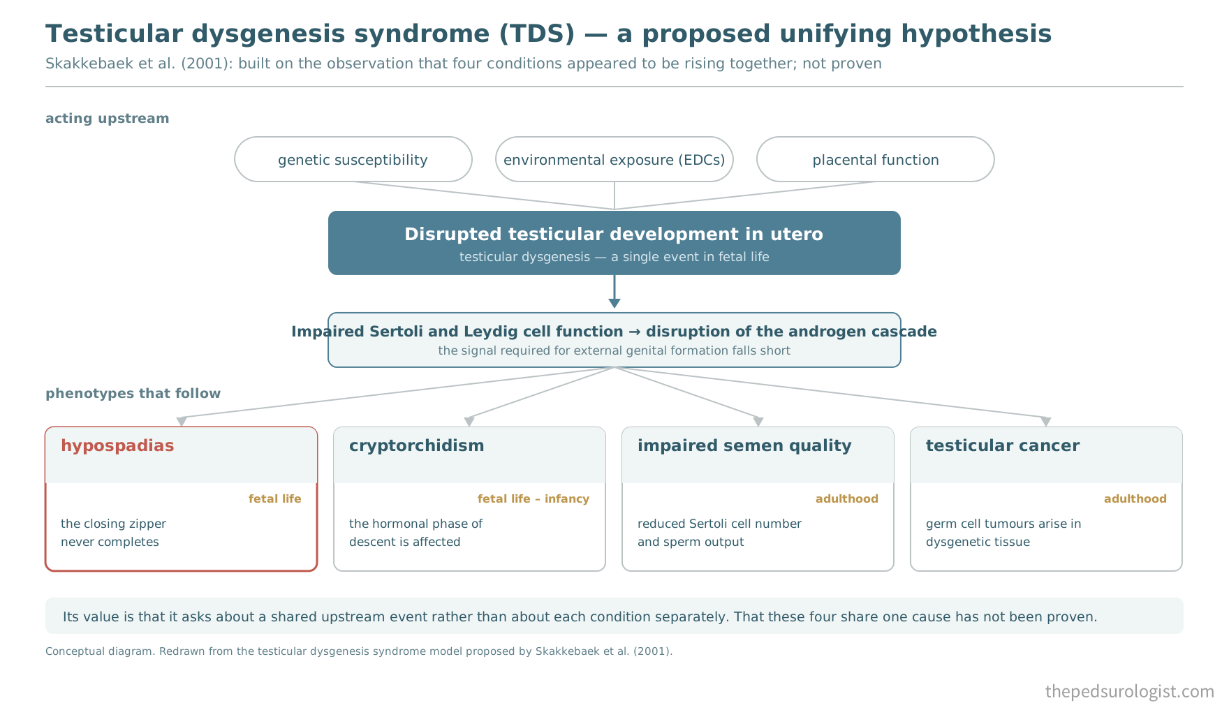
The hypothesis is useful because it makes one ask about a shared upstream event rather than interrogating each cause separately. Looking for the cause of hypospadias is not separable from looking for the cause of cryptorchidism, infertility and testicular cancer. It remains a hypothesis: that these four really arise from one cause has not been proven.
Placental insufficiency — the most consistent association
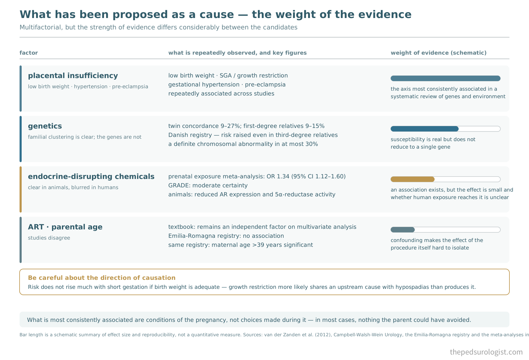
A systematic review covering both genes and environment (van der Zanden et al., 2012) concluded that what was consistently associated was low birth weight, gestational hypertension and pre-eclampsia. The authors read this as pointing to a common axis of placental insufficiency: reduced placental hCG secretion lowers androgen production by the fetal testis, and the closing zipper does not complete.
Low birth weight, small-for-gestational-age growth or intrauterine growth restriction, gestational hypertension and pre-eclampsia have been associated repeatedly across studies, and together they point to placental dysfunction as a common axis. Direct measurement of the placenta runs the same way: low placental weight at birth has been reported to carry roughly a twofold risk of hypospadias and about 1.5-fold for cryptorchidism. Reports that prematurity and low birth weight cluster more strongly where proximal hypospadias and cryptorchidism occur together point to the same axis.
Whether low birth weight caused the hypospadias, or whether the two share a cause, is a different question. Risk rises with the degree of smallness for gestational age, but analyses show that short gestation with adequate weight does not raise risk much. That suggests growth restriction did not produce the hypospadias so much as both diverged from the same upstream cause. When the placenta underperforms, the baby is small and androgen is short — neither one produced the other.
Endocrine-disrupting chemicals — the gap between animals and humans
The animal evidence is clear. Exposure to phthalates such as DBP and DEHP reduces androgen receptor expression and 5α-reductase activity and produces hypospadias, and the mechanism is reasonably well worked out.
In humans the picture changes. A meta-analysis of prenatal exposure and hypospadias reported an odds ratio of 1.34 (95% CI 1.12–1.60) with moderate certainty on GRADE assessment, the association being strongest for organochlorines and glycol ethers. An association exists, but the effect size does not compare with the placental axis.
The textbook is blunter: in animals a direct link between pesticide exposure, disruption of the androgen cascade and external genital anomalies has been established, but attempts to establish that link in humans have not been successful. A case-control study from southern France did find significant risk from parental exposure to detergents, pesticides and cosmetics during pregnancy, with higher risk where occupational exposure was added. These compounds are everywhere, which makes isolating any one of them difficult.
The 2012 systematic review's formulation still holds — exogenous endocrine disruptors have the potential to cause hypospadias, but whether actual human exposure reaches the level needed to produce that effect is unclear. And as noted above, the difference between the animal and human mechanisms of urethral formation widens the gap further.
If exposure is hard to measure, its consequence can be. Anogenital distance (AGD), measured from the centre of the anus to the penoscrotal junction, is used as a marker of fetal androgen action. AGD is shorter in populations with suspected endocrine disruptor exposure, short AGD is associated with infertility, cryptorchidism and hypospadias, and it correlates with the severity of hypospadias. That a value obtainable with a tape measure in clinic reflects the androgen environment of fetal life is a striking thing.
Assisted reproduction and parental age — inconsistent
Reports of increased hypospadias after assisted reproductive technology have appeared for years, but studies disagree. In the Emilia-Romagna dataset cited above there was no association with ART, with paternal age or with maternal body mass index. In the same dataset, maternal age over 39, multiple pregnancy, prematurity and small-for-gestational-age weight were all significantly associated.
The textbook, by contrast, states that ART remains an independent factor on multivariate analysis — the association persisting after adjustment for the higher frequency of prematurity, low birth weight and multiple pregnancy.
Interpretation is difficult because of confounding. Parents undergoing ART are older, have more multiple pregnancies, and have higher rates of prematurity and low birth weight. And subfertility itself may reflect something about parental reproductive function — within the TDS framework, a father's reduced semen quality and his son's hypospadias may share an upstream cause. Separating the effect of the procedure from the effect of the condition that led to it is not easy.
Genetics — much found, little explained
At the single-gene level, variants in WT1, SF1, BMP4, BMP7, FGF8, FGFR2, AR, HSD3B2, SRD5A2, ATF3, MAMLD1, MID1 and BNC2 have been reported; at the polymorphism level HSD17B3, ESR1, ESR2, DGKK, CYP1A1, GSTM1 and GSTT1 are added.
Familial clustering is clear. Concordance among twins, monozygotic and dizygotic together, is 9–27%, and the probability that a patient has a first-degree relative (brother or father) with the same condition is 9–15%. A Danish registry study following more than a million individuals confirmed both maternal and paternal transmission, with risk elevated even in third-degree relatives.
Yet attempts to pin down the genes have not delivered what was hoped. A definite chromosomal abnormality is identified in at most 30%. Nearly 200 syndromes with a known genetic cause include hypospadias, but these account for only a fraction of cases. A clear genetic predisposition that does not reduce to a single gene — a mixture of monogenic and multifactorial forms — is where the understanding currently stands.
So what do we tell the parent?
Back to that question — whether there was something the parent did during the pregnancy.
The honest answer is that in most cases the cause cannot be identified, and it was not something the parent could have avoided. What has been most consistently associated is a set of conditions relating to placental function, and those are conditions of the pregnancy rather than choices made during it. Endocrine disruptors are associated but with a small effect size; assisted reproduction gives conflicting results.
There is a separate reason why this explanation matters. Saying the cause is unknown makes a parent anxious; saying the cause cannot be identified releases them from blame. The same fact, delivered in two different directions, does two different things.
And one thing can be added. Where hypospadias occurs with a non-palpable testis, a disorder of sex development must be excluded, so separate investigation immediately after birth is recommended. If the answer to "why" is not to be merely "we don't know", then what needs to be checked now should at least be said clearly.
In the next instalment
Having seen the two steps of urethral formation in Part 4, the next instalment digs into one point within them. Why does the penis curve?
The clue is already on the table. In a classic analysis of 46 cases, chordee was present in all 7 perineal and scrotal cases, 15 of 16 penile and coronal cases, and 11 of 23 glanular cases. Frequency tracks severity, but it is present in nearly half even of the mildest forms. And the mechanism has been proposed to differ by severity — in perineal forms, absence of androgen stimulation leads to apoptosis of the urethral plate and the corpora themselves bend, whereas distally it is less a bending of the corpora than deficient periurethral growth that produces the curvature.
Hypoplasia of the ventral tissues, tethering by the urethral plate and the tissue beneath it, or asymmetry of the corpora themselves — authors differ, and that difference translates directly into a difference in operative strategy. Part 6 takes up the root of why Part 1 placed curvature at the centre of this series.
References
- Yu X, Nassar N, Mastroiacovo P, et al. Hypospadias prevalence and trends in international birth defect surveillance systems, 1980-2010. Eur Urol. 2019;76(4):482-490.
- Ballardini E, Armaroli A, Finessi N, Maietti E, Astolfi G, Neville AJ. Hypospadias prevalence in the Emilia Romagna Region registry: increasing or methodology? J Pediatr Urol. 2020;16(4):448.e1-448.e7.
- Fichtner J, Filipas D, Mottrie AM, Voges GE, Hohenfellner R. Analysis of meatal location in 500 men: wide variation questions the need for meatal advancement in all pediatric anterior hypospadias cases. J Urol. 1995;154(2 Pt 2):833-834.
- Paulozzi LJ, Erickson JD, Jackson RJ. Hypospadias trends in two US surveillance systems. Pediatrics. 1997;100(5):831-834.
- Dolk H. Epidemiology of hypospadias. In: Hadidi AT, Azmy AF, eds. Hypospadias Surgery: An Illustrated Guide. Berlin: Springer; 2004:51-56.
- Shan X, Aguilar S, Canon S. Hypospadias prevalence by severity and associated risk factors: a population-based active surveillance analysis. J Pediatr Urol. 2023;19(6):720.e1-720.e9.
- Nordenvall AS, Frisén L, Nordenström A, Lichtenstein P, Nordenskjöld A. Population based nationwide study of hypospadias in Sweden, 1973 to 2009: incidence and risk factors. J Urol. 2014;191(3):783-789.
- Skakkebaek NE, Rajpert-De Meyts E, Main KM. Testicular dysgenesis syndrome: an increasingly common developmental disorder with environmental aspects. Hum Reprod. 2001;16(5):972-978.
- van der Zanden LFM, van Rooij IALM, Feitz WFJ, Franke B, Knoers NVAM, Roeleveld N. Aetiology of hypospadias: a systematic review of genes and environment. Hum Reprod Update. 2012;18(3):260-283.
- Kalfa N, Paris F, Philibert P, et al. Is hypospadias associated with prenatal exposure to endocrine disruptors? A French collaborative controlled study of a cohort of 300 consecutive children without genetic defect. Eur Urol. 2015;68(6):1023-1030.
- Schnack TH, Zdravkovic S, Myrup C, et al. Familial aggregation of hypospadias: a cohort study. Am J Epidemiol. 2008;167(3):251-256.
- Cook WA, Stephens FD. Pathoembryology of the urinary tract. In: King LR, ed. Urological Surgery in Neonates and Young Infants. Philadelphia: Saunders; 1988:1-23.
- Hypospadias. In: Partin AW, Dmochowski RR, Kavoussi LR, Peters CA, eds. Campbell-Walsh-Wein Urology. 13th ed. Philadelphia: Elsevier. (Chapter 56)
- EAU Guidelines on Paediatric Urology. Hypospadias. European Association of Urology, 2026.