The question we are asked most often in clinic contains two questions. How likely is this to go away on its own, and is it safe to wait while it does? We usually answer both with the grade — but the scale that answer rests on is softer than it looks, and it does not answer the two questions with equal accuracy.
This article looks at what spontaneous resolution actually is structurally, how well the grade knows it, and what can be placed alongside the grade.
Resolution is not the crossing of a single number
That reflux may disappear as a child grows is well known. But explaining it only as "the ureter grows" leaves out the important part. The ureterovesical junction is not a fixed tube. Its shape and the pressures around it change with every cycle of filling and emptying, and the ureter opens and closes inside that. Resolution has to be read as geometric growth, maturation of bladder function, and remodelling of the supporting tissue and the ureteral wall, together.
Scroll sideways to see the full figure
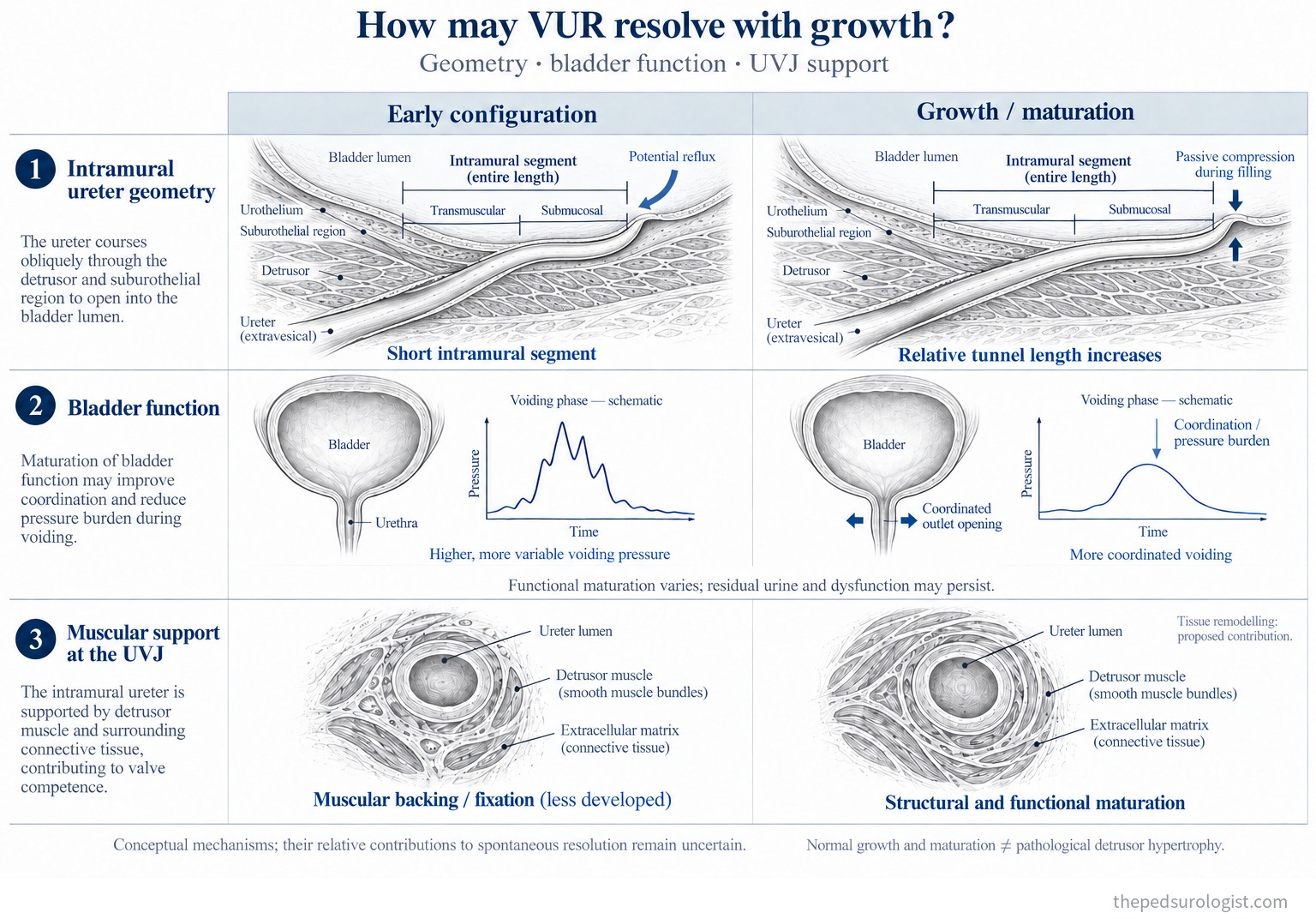
First, the closing segment lengthens relative to the calibre of the ureter. What matters is not length in absolute terms. For the same length, the closure a segment provides depends on the diameter of the ureter: a short wide channel and a long narrow one are not the same structure. Paquin's data in normal children, seen in Part 1, sits behind this — intravesical and submucosal length rise with age while the increase in diameter is comparatively small. But that is an anatomical observation in normal children, not a record of a tunnel growing in a refluxing child until reflux disappears. It is a reasonable explanation, not a formula, and in particular 5:1 cannot be used as a threshold for spontaneous resolution.
Second, the load the bladder places on the junction changes. Here storage pressure and voiding pressure must be kept apart. When "bladder pressure falls" is offered as an explanation for improvement without saying which one, two different phenomena get bundled together. Infants may show high voiding pressures and immature coordination; as voiding becomes more efficient with growth, the abnormal load on the junction falls. But not every bladder normalises with age — in infants followed with high-grade reflux, initially high voiding pressures returned to the normal range, yet some were left with incomplete emptying, large capacities and residual urine.
One more distinction. The pressure the bladder generates and the pressure at which reflux begins are different things. The first is the load on the junction; the second reflects how much the junction holds against. The finding below — that reflux appearing at larger filling volumes resolves more often — belongs to the second, and does not mean high bladder pressure is beneficial.
Third, support and the way it works. For the intramural ureter to be compressed shut, it needs tissue backing it and a structure holding it in place. Adequate length will not work the same way over weak backing or insufficient fixation. But "the detrusor hypertrophies and the reflux stops" is a formulation to avoid. Muscular development with normal growth and pathological hypertrophy in response to obstruction or high pressure are not the same thing, and a thicker wall is not in itself better anti-reflux function. What matters is not thickness but arrangement, backing, fixation and coordination.
The reach of the evidence. Histological studies of refluxing distal ureters show smooth-muscle atrophy and disarray, collagen accumulation and extracellular-matrix remodelling, so tissue remodelling is also proposed as a mechanism of resolution. But remodelling seen in ureters that came to surgery is not evidence that the same change abolished reflux in ureters that resolved on their own. It widens the mechanism usefully; it is not established as a cause.
Resolution, then, is not explained by "the moment the tunnel reaches 5:1" or "the moment bladder pressure drops below some value." The outcome is unambiguous; the route to it differs between children. That is why a single grade predicts the natural history poorly, and why ureteral calibre and bladder function belong in the same look.
Resolution rates by grade
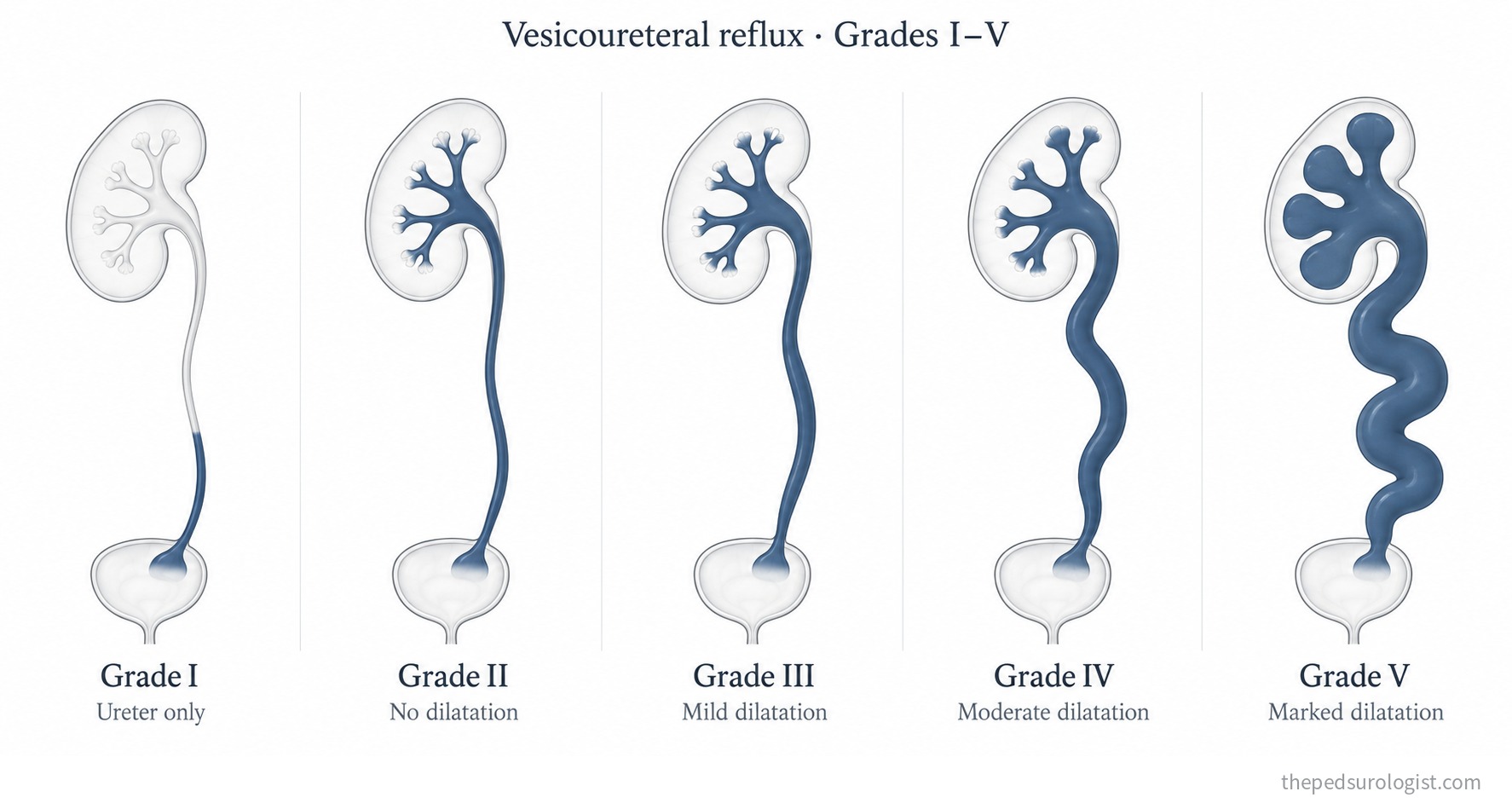
The 1981 International Reflux Study Committee classification is still in use. It helps to see what separates the five categories before taking on the numbers.
Scroll sideways to see the full figure
| Grade | Appearance | 2-year resolution Estrada nomogram | What to read alongside it |
|---|---|---|---|
| I | Into a non-dilated ureter only | 72% | Most resolve. Age and infection history govern management more than the grade itself |
| II | To the pelvis and calyces, without dilatation | 61% | The most scattered figure across studies, 63–85%. A grade II with a notably wide distal ureter may not behave like a typical grade II |
| III | Mild to moderate dilatation of ureter, pelvis and calyces; minimal blunting of the fornices | 49% | About half. Of those that resolved, 92% did so within four years in one report |
| IV | Moderate ureteral tortuosity with pelvicalyceal dilatation | 32% IV–V combined | Spontaneous resolution is uncommon. In infants with high-grade reflux, 29% resolved in the first year, falling to 9% per year thereafter. Note too that the IV-versus-V boundary is the least stable one — section 3 |
| V | Gross dilatation, loss of papillary impressions, marked ureteral tortuosity |
Resolution broadly runs inverse to the initial grade. But the figures above come from one cohort and one estimate, and they shift when set beside other studies.
There are reasons for that. Follow-up differs, what counts as resolution differs (some studies count improvement to grade 0–II), and above all, when a larger share of the high-risk group goes to surgery, the data that remain under follow-up tilt toward optimism.
How solid is that grade?
The grade is assigned by eye, from a dynamic study. A voiding cystourethrogram is not one picture but a sequence of filling and voiding, and what is seen depends on which frame is taken as representative. That variability was quantified in the RIVUR cohort. Schaeffer and colleagues compared enrolment studies read by one local radiologist and, independently, by two blinded reference radiologists — 602 reports from 90 institutions, 560 left and 524 right ureters.
- All three agreed on the grade in 59% of ureters; by participant, with both ureters concordant, in 43%.
- The local read differed from the adjudicated reference grade by exactly one grade in 19% of ureters, and by two or more in 2.2%.
- By contrast, agreement on other findings — whether bladder shape was normal, for instance — was 92% or better. The reading is not unstable; the grade, specifically, is.
The study was limited to children with a history of urinary tract infection and grade I–IV reflux, so it does not generalise to every cystogram. It is, however, exactly the population in whom grade drives treatment decisions.
"Considering that any reflux grade is assigned based on a dynamic voiding study, a variation of at least one grade, particularly when assigning a higher grade, is quite realistic. Therefore, attempting to discriminate true differences in resolution rates for VUR of grades III and higher may not be particularly clinically relevant."
This is not an argument for abandoning the grade but for using it at its true resolution. It is adequate to separate I–II from IV–V. It should not carry a decision that turns on IV versus V, and where that single step would change management, something beyond the grade has to be in hand.
What the grade misses is equally clear. The classification is driven by deformity of the collecting system, and the ureter enters only at grade III, and only through the words "dilatation" and "tortuosity." So reflux with a substantially dilated distal ureter but an almost undistorted collecting system is, read literally, grade II. Campbell illustrates this case separately and notes it may not behave like a typical grade II system.
The ureteral diameter ratio
The measure proposed by Cooper and colleagues captures that missing variable as a number.
Scroll sideways to see the full figure
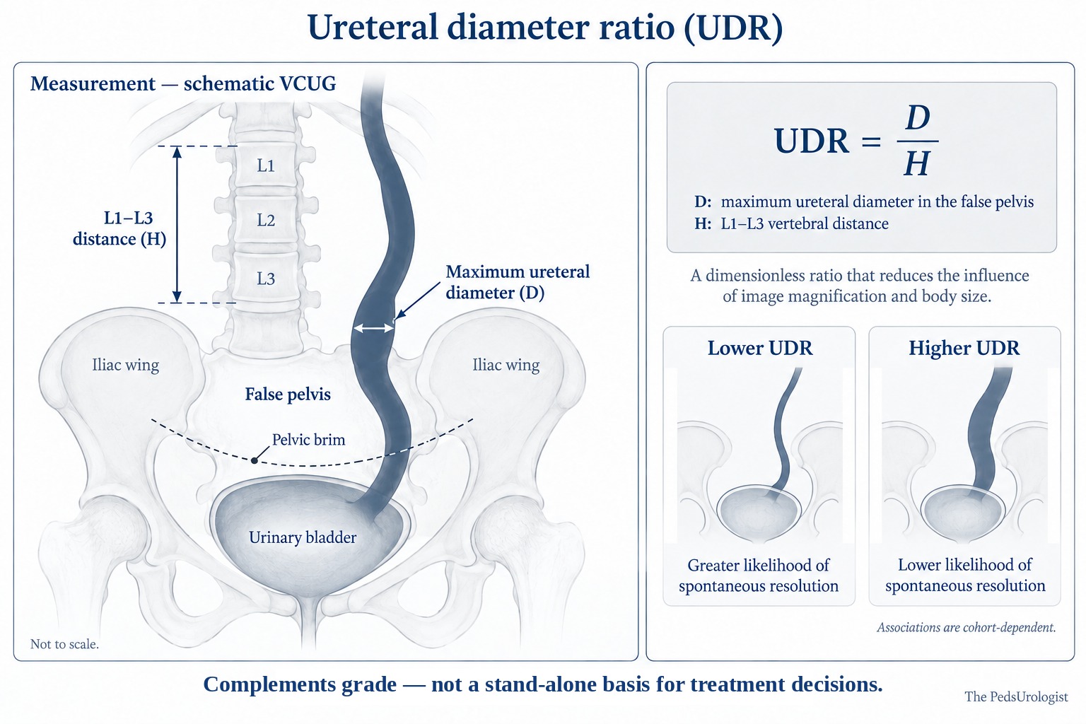
- Early spontaneous resolution — in 147 children diagnosed at a mean of 5.5 months, grade (p=0.001) and UDR (p<0.0001) both entered the multivariable model significantly. Odds of persistence rose 2.6-fold per 0.1 increase in UDR versus 1.6 per grade (not significant), and no child above 0.43 resolved early.
- Resolution and endoscopic injection — in 383 patients, UDR outperformed grade for predicting resolution (p<0.001), and each 0.05 increase reduced the success of injection. But 84% of that cohort underwent surgery, so the resolution rate itself cannot be generalised — the bias described in section 2.
- Breakthrough febrile UTI — in 140 children, UDR was 0.36 versus 0.25, and controlling for grade, each 0.1 increase carried 1.7 times the odds.
So the place of UDR is beside the grade, not instead of it — the evidence being that grade remained significant in the same model. Where the grade describes how damaged the collecting system looks, UDR describes how wide the ureter is immediately above the junction, and being a continuous measurement it depends far less on reader interpretation.
Which variable predicts what
The same variable gives a different answer when the question changes. Split by the two questions:
| Variable | Predicts resolution? | Predicts infection risk while waiting? |
|---|---|---|
| Reflux grade | Yes, though a one-grade difference above III is not trustworthy | Weakly. Did not reach significance for breakthrough febrile UTI (p=0.09) |
| UDR | Yes, entering the model alongside grade | Yes, independently of grade |
| VUR Index | Yes; stratifies best | Yes; highest AUC of the three (0.77) |
| Age at diagnosis | Yes; under 12 months independently significant (p=0.008) | Indirectly — infancy is the high-risk window for febrile infection |
| Filling volume at onset of reflux | Yes; onset beyond 75% of predicted capacity favours resolution (p=0.0005) | Little direct data |
| Bladder–bowel dysfunction | A condition that impedes resolution | Yes; an independent variable that imaging does not substitute for |
| Prior febrile urinary tract infection | A negative predictor (p<0.0001) | Yes; where the two questions interlock |
| Sex | Varies by age group; in children diagnosed after 24 months, female sex predicted non-resolution (p=0.005) | Hard to use on its own |
| Associated ureteral anomalies complete duplication · periureteral diverticulum | Yes; associated with non-resolution in the first 12–18 months (HR 0.29) | Little direct data |
What to read in the table is not the ranking but the first row. When Arlen and colleagues followed 139 children diagnosed at a mean of 5.4 months for a mean of 32 months, with breakthrough febrile urinary tract infection as the outcome, the VUR Index (AUC 0.77) and UDR (0.71) were significant while grade did not reach significance (p=0.09, AUC 0.68). A variable that served well for predicting resolution loses its force when the question becomes infection risk. That is why the two questions should not be answered with one measure.
Three additions.
The VUR Index sums sex, grade, ureteral anomalies and the timing of reflux into a 1–6 point score. In a multi-institutional validation of 369 children diagnosed under 24 months, improvement or resolution ran 88.2% · 77.3% · 62.3% · 32.1% · 14.3% for scores of 1, 2, 3, 4 and 5 or more, with median times to resolution of 12.6, 12.7, 15.1, 25.6 and 31 months or longer.
What "ureteral anomalies" means in that table is not a loose category. In the study that derived the index, the items entered into the multivariable analysis were complete ureteral duplication and periureteral (Hutch) diverticulum — the same anatomical abnormalities whose classification was disputed in Part 1 now reappearing as prognostic variables. Ureterocele is not part of that definition, as studies of primary reflux commonly exclude it; the natural history of reflux with a ureterocele cannot be inferred from these figures and is better treated as a separate problem.
Finally, bladder–bowel dysfunction showed no significant correlation with UDR (Payza, p>0.05): imaging will not report it, so constipation and voiding habits have to be asked about separately.
Observation is a plan
Following the logic of section 1, the conclusion is plain. Resolution is a process of junctional remodelling and stabilising bladder mechanics, so observation is an active plan to create the conditions for that process. What is actually being agreed when we say "let's wait" is three things.
- A response to infection. Decide in advance where and what will be checked when there is a fever, and whether antibiotic prophylaxis will be used. The evidence on prophylaxis is substantial enough for its own Evidence note, but whichever way that decision goes, observation without a plan for fever is not observation.
- Management of bladder and bowel function. In the mechanism of section 1 it is a condition of resolution; in the data of section 5 it is an independent variable for infection risk. It is the rare place where one intervention acts on both questions.
- The interval and the stopping rule for follow-up. Write down at the outset when imaging will be repeated and what event would change the plan. The fact from section 2 — that the annual probability of resolution falls year on year — is what justifies not repeating follow-up indefinitely.
Campbell's section on predictive tools closes with this sentence.
"Because the greatest threat to a patient with reflux is the consequence of infection, the ability to predict who is at higher risk for UTI rather than predict VUR persistence or resolution may be more clinically relevant."
That is where Part 1 arrived as well. What we actually want to know is not when the reflux will disappear but whether this child's kidney will be harmed. Predicting resolution was only a surrogate for that question — and on the side that matters more, our instruments are weaker.
References
- Khoury AE, Wehbi E. Management Strategies for Vesicoureteral Reflux. In: Campbell-Walsh-Wein Urology, 13th ed. Elsevier.
- Lebowitz RL, Olbing H, Parkkulainen KV, et al. International system of radiographic grading of vesicoureteric reflux. Pediatr Radiol. 1985;15(2):105-109.
- Schaeffer AJ, Greenfield SP, Ivanova A, et al. Reliability of grading of vesicoureteral reflux and other findings on voiding cystourethrography. J Pediatr Urol. 2017;13(2):192-198. doi:10.1016/j.jpurol.2016.06.020
- Cooper CS, Alexander SE, Kieran K, Storm DW. Utility of the distal ureteral diameter on VCUG for grading VUR. J Pediatr Urol. 2015;11(4):183.e1-6.
- Arlen AM, Kirsch AJ, Leong T, Cooper CS. Validation of the ureteral diameter ratio for predicting early spontaneous resolution of primary vesicoureteral reflux. J Pediatr Urol. 2017;13(4):383.e1-383.e6. doi:10.1016/j.jpurol.2017.01.012
- Arlen AM, Leong T, Guidos PJ, Alexander SE, Cooper CS. Distal ureteral diameter ratio is predictive of breakthrough febrile urinary tract infection. J Urol. 2017;198(6):1418-1423. doi:10.1016/j.juro.2017.06.095
- Payza AD, Hoşgör M, Serdaroğlu E, Sencan A. Can distal ureteral diameter measurement predict primary vesicoureteral reflux clinical outcome and success of endoscopic injection? J Pediatr Urol. 2019;15(5):515.e1-515.e8. doi:10.1016/j.jpurol.2019.07.005
- Arlen AM, Leong T, Wu CQ, Traore EJ, Cooper CS, Kirsch AJ. Predicting breakthrough urinary tract infection: comparative analysis of vesicoureteral reflux index, reflux grade and ureteral diameter ratio. J Urol. 2020;204(3):572-577. doi:10.1097/JU.0000000000001035
- Kirsch AJ, Arlen AM, Leong T, et al. Vesicoureteral reflux index (VURx): a novel tool to predict primary reflux improvement and resolution in children less than 2 years of age. J Pediatr Urol. 2014;10(6):1249-1254. doi:10.1016/j.jpurol.2014.06.019
- Arlen AM, Garcia-Roig M, Weiss AD, Leong T, Cooper CS, Kirsch AJ. Vesicoureteral reflux index: 2-institution analysis and validation. J Urol. 2016;195(4 Pt 2):1294-1299. doi:10.1016/j.juro.2015.03.094
- Garcia-Roig M, Ridley DE, McCracken C, Arlen AM, Cooper CS, Kirsch AJ. Vesicoureteral reflux index: predicting primary vesicoureteral reflux resolution in children diagnosed after age 24 months. J Urol. 2017;197(4):1150-1157. doi:10.1016/j.juro.2016.12.008
- McMillan ZM, Austin JC, Knudson MJ, Hawtrey CE, Cooper CS. Bladder volume at onset of reflux on initial cystogram predicts spontaneous resolution. J Urol. 2006;176(4 Pt 2):1838-1841. doi:10.1016/S0022-5347(06)00619-7
- Estrada CR Jr, Passerotti CC, Graham DA, et al. Nomograms for predicting annual resolution rate of primary vesicoureteral reflux. J Urol. 2009;182(4):1535-1541.
- Individual resolution figures by grade (McLorie, Cha and Lee, Sjöström, Wildbrett), the follow-up of infant bladder function, and the histology of refluxing ureters follow reports cited in Chapter 41 of Campbell-Walsh-Wein. Literature searching used PubMed.