
When the urinary infection has been treated and the reflux has gone, questions about the kidney remain. Will a kidney that was small from the start grow as the child grows? Will a scar left after infection stay as it is, or lead over time to declining function? To parents these are all one question — "Will the kidney be all right?" — but answering it means looking at how much functioning tissue remains now and at how things develop from there.
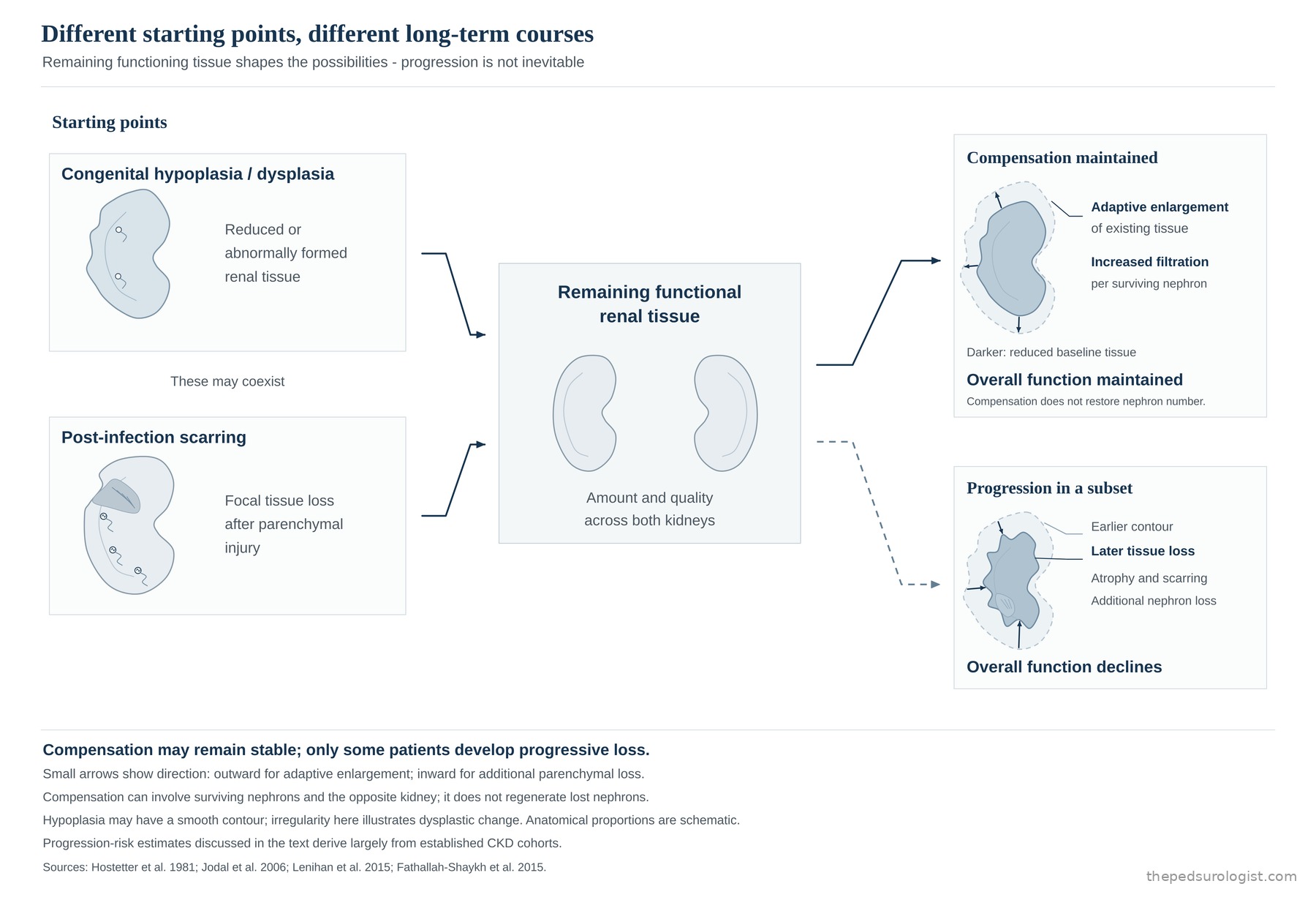
Congenital hypodysplasia and post-infectious scarring start from different places. Over the long term, though, both lead to the same question: can the renal tissue that remains sustain enough function through growth and time?
The same reflux, different starting kidneys
In congenital hypoplasia the number of nephrons may be reduced from the outset; in dysplasia the differentiation and architecture of the tissue itself form abnormally. A post-infectious scar is the result of part of the tissue injured by pyelonephritis failing to recover and becoming fibrotic. Both processes may coexist in one child, and without imaging from before the infection it can be hard to assign a parenchymal abnormality found later clearly to either.
Judging long-term prognosis therefore means looking at the renal parenchyma alongside the grade of reflux. What matters is whether the lesion is confined to one side or present on both, whether the contralateral kidney has developed adequately, and at what level total renal function starts. The future of a child with high-grade reflux and preserved bilateral function cannot be described with the same data as that of a child whose function is already reduced by bilateral hypodysplasia.
The same distinction is needed when reading the long-term data cited here. Much of what the literature calls natural history is a course observed under treatment and regular follow-up, and many studies did not clearly separate congenital dysplasia from acquired scarring. In particular, most of the paediatric data that put a number on the risk of progression come from populations that already had chronic kidney disease. Those rates cannot be transferred to a child with normal total function and only a focal scar.
Scroll sideways to view
A small kidney does not necessarily keep getting worse
When functioning tissue is lost, the remaining nephrons and the contralateral kidney take over part of the work. As the remaining tissue enlarges and each nephron filters more, total renal function can be sustained for a long time even with a small kidney on one side or a focal scar. This growth does not mean lost nephrons have been replaced. It is an adaptation in which the remaining tissue carries more of the load.
The same adaptation can be seen directly in people. In a study following 21 adult living kidney donors for a median of six years after donation, GFR in the remaining kidney rose by about 40%, explained by hypertrophy and increased perfusion of the remaining glomeruli, with no evidence of glomerular hypertension. It was a small study, and hypertension became more frequent among the donors over the same period.
Some children do follow a relatively stable course on long-term follow-up. In the ten-year results of the International Reflux Study reported by Jodal and colleagues, children with grade III–IV reflux who entered with a GFR of at least 70 mL/min/1.73 m² generally kept good renal function under both medical and surgical management. New scars appeared mostly within the first five years and very rarely thereafter, and among the children followed, only one had a GFR that fell below 70.
These results were observed under close supervision and prompt treatment of recurrent infection, and cannot be applied as they stand to children whose function was already reduced at the outset. They nonetheless show that high-grade reflux by itself should not be taken to predict progressive loss of function.
The IRS randomised 306 children under 11 years with non-obstructive grade III–IV reflux, previous UTI and GFR ≥70 to antibiotic prophylaxis or ureteral reimplantation. Up to five years there were 40 new scars (19 medical, 21 surgical); between five and ten years, two more. Ten-year follow-up was available for 252 children and follow-up imaging for 223, with scars assessed by urography. Three children developed hypertension requiring treatment.
In some, function declines even without infection
Compensation is not always sufficient or permanent. To maintain total filtration with fewer functioning nephrons, each remaining glomerulus must handle more blood flow and more filtration. Raised intraglomerular pressure and single-nephron GFR are at first an adaptation that preserves function, but if the load is sustained it can lead to glomerular injury and sclerosis. Functioning nephrons then fall further, and the burden on those remaining rises again.
The hyperfiltration model proposed by Hostetter, Brenner and colleagues from renal ablation experiments is an important framework for this progression. Single-nephron GFR rose in the remnant glomeruli, accompanied by structural glomerular injury. In animals given a low-protein diet that prevented the rise in filtration, that injury was largely attenuated.
The experiment used rats with one kidney removed and five-sixths of the other infarcted. The scale of tissue loss bears no comparison with a child who has one small kidney or a focal scar. It is a framework for understanding mechanism, not evidence that every hypoplastic kidney or focal scar follows this path.
Injury that begins in the glomerulus does not stay there. As sclerosed glomeruli accumulate and proteinuria appears, inflammation and fibrosis extend into the tubules and interstitium. This tubulointerstitial injury is common to chronic kidney disease whichever compartment the disease began in, and it is the degree of tubulointerstitial fibrosis, more than glomerular injury, that tracks the decline in function. In kidneys removed from patients who had reached end-stage disease from reflux nephropathy, FSGS (focal segmental glomerulosclerosis) was very common and accounted for their proteinuria.
What matters clinically is that laboratory values can stay normal for a long time while this is under way. While the remaining nephrons raise their filtration to hold total GFR, creatinine barely moves even as nephrons are being lost. Decline becomes visible only once the reserve is exhausted, and the smaller the remaining mass, the larger the share each further loss represents, so the fall can appear to accelerate. The turn in the kidney-survival curve around puberty in the ItalKid data in section 5 can be read in this light, although the authors did not identify its mechanism. Nor does every child follow this course; in CKiD, for example, the average decline was gradual. That is why a normal GFR alone is no reason for reassurance; proteinuria, blood pressure and renal growth need to be watched alongside it.
This perspective explains why, in some patients, function can decline even after the reflux has resolved and infection has not recurred: there is a course of kidney disease that proceeds from an already reduced mass of functioning tissue. Subsequent decline therefore cannot all be attributed to new infection or persisting reflux. The amount of tissue that remained at the start, blood pressure and proteinuria, and further infection or lower urinary tract dysfunction must all be considered together.
Loss on one side and loss of total function are different things
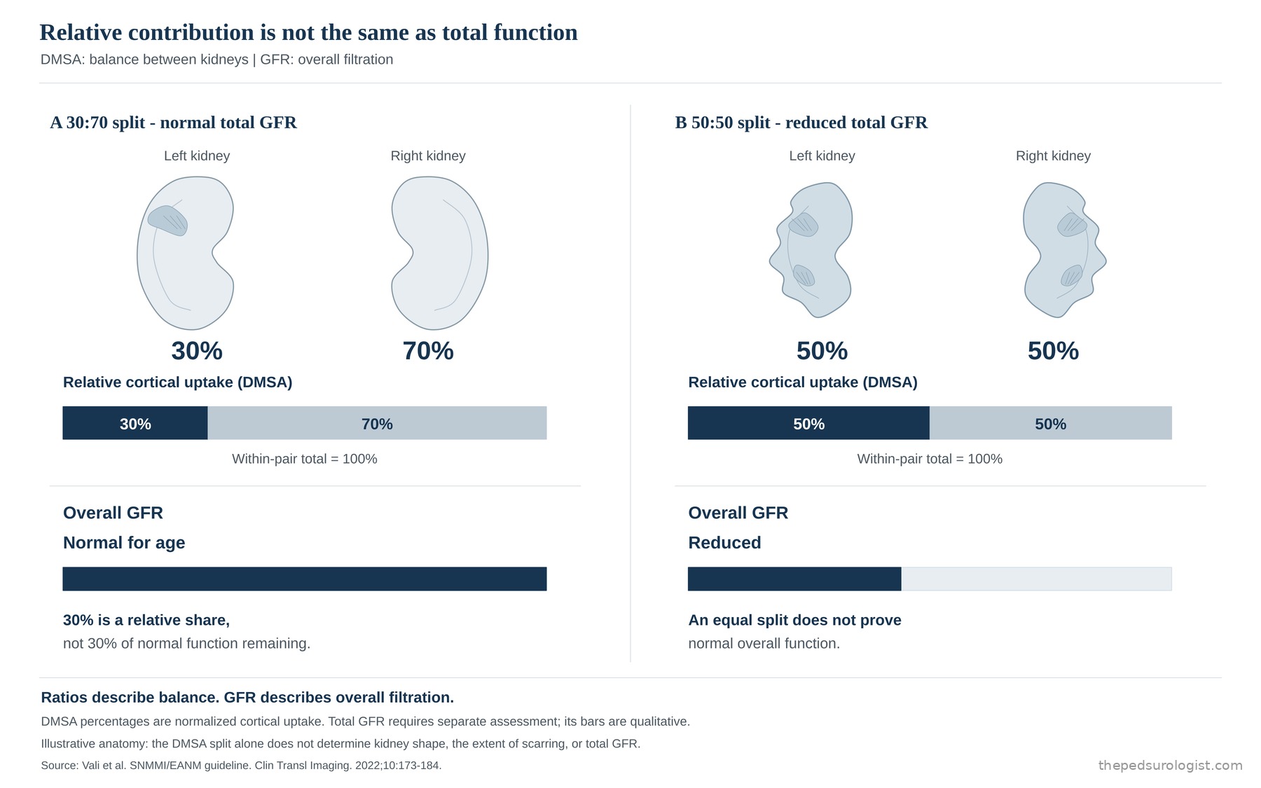
If DMSA reports split function as 30:70, that describes the relative share of cortical uptake between the two kidneys. It does not mean the kidney retains only 30% of normal function. Conversely, a 50:50 split is no guarantee that total function is normal, since both kidneys may be similarly reduced. Split function assesses the balance between the two sides; GFR assesses total filtration.
Scroll sideways to view
In long-term data, bilateral parenchymal abnormality is likewise an important dividing line. When Lahdes-Vasama and colleagues assessed patients treated for reflux in childhood at a mean age of 41, reduced function, proteinuria and hypertension were more prominent in those with bilateral scars. GFR was below normal in 83% of the bilateral group, mostly mildly, and the four patients with moderate or worse impairment all had bilateral scars. Those with unilateral scars were broadly similar to those without, but tended towards higher blood pressure, so blood pressure warranted attention even with a scar on one side.
Scarring in this study was assessed by ultrasound, not DMSA, and in 42% of participants no scar was seen on ultrasound. Of the original 267 patients, renal-function information was available for 147; the 12 who had died of kidney-related causes and the 8 who had reached end-stage renal failure were not among the 127 analysed. The proportions among those 127 therefore cannot be taken to represent the full disease burden of the original cohort. At the same time, the status of the remaining patients without information is unknown, so the direction of selection bias cannot be fixed either. As data from a historical treated cohort, the proportions should not be transferred to the risk of children diagnosed with VUR today.
Reading the lesion on imaging alongside the change happening now
Long-term follow-up depends not only on renal size and cortical appearance but on blood pressure, persistent proteinuria and the trend in eGFR. Even where the extent of scarring looks unchanged on imaging, new or increasing proteinuria, rising blood pressure or falling eGFR call for the current course to be reassessed. This is why change in renal function adjusted for growth and body size should be read rather than a single creatinine value compared.
Data from children who already have chronic kidney disease indicate what to look for. In the ItalKid study of Ardissino and colleagues, among children with reduced function from hypodysplastic nephropathy, greater proteinuria was followed by faster decline. In the CKiD study of Fathallah-Shaykh and colleagues, proteinuria in children with nonglomerular CKD was likewise associated with faster GFR decline. Proteinuria was consistent across both studies.
The blood-pressure findings differed by study. In CKiD, systolic blood pressure was associated with faster GFR decline independently of proteinuria, whereas ItalKid found no association with mean arterial pressure. The two studies differed in population and in blood-pressure measure, so rather than one refuting the other, the accurate reading is that proteinuria was a consistent predictor, while whether an association with blood pressure was found differed between studies.
ItalKid (2004) followed 225 children with hypodysplasia and creatinine clearance of 20–75 mL/min/1.73 m² for a mean of 3.5 years, and to examine the relationship between proteinuria and progression it excluded patients receiving ACE inhibitors. CKiD followed 522 children with nonglomerular CKD (mean baseline GFR 52) for a median of 4.4 years, during which GFR fell by an average of 1.3 mL/min/1.73 m² per year.
During growth the trend needs to be checked again. In ItalKid data following 935 children with CKD from hypodysplasia, the kidney-survival curve was not linear but turned steeply around puberty, with the break-point at 11.6 years in boys and 10.9 years in girls. This should not be read to mean that every scarred kidney worsens at puberty. It means that in a child whose function is already reduced, stable results in early childhood do not settle the later course.
There is still work to do after the reflux has resolved
Correcting reflux addresses the problem at the ureterovesical junction and, in some patients, is an important treatment for reducing febrile urinary tract infection. It does not reverse hypodysplasia or scarring already present. In the International Reflux Study, the group treated with antibiotic prophylaxis had more febrile infections than the group that underwent ureteral reimplantation, yet new scars were similar in number (19 vs 21) and no difference was found between the groups in renal growth, radionuclide imaging or renal function. Within that study's population and conditions of follow-up, reducing infection and long-term renal outcome were different endpoints. Judging the indication for surgery and assessing the long-term prognosis of the remaining kidney are both needed, separately.
Nor is subsequent management simply a matter of waiting for deterioration. In the ESCAPE trial of children with chronic kidney disease, with every child receiving an ACE inhibitor, the group assigned a lower blood-pressure target had less deterioration of renal function. This does not mean every child with VUR needs the same drug treatment, but it shows that the course of a child with established kidney disease can be influenced. This is why bilateral parenchymal abnormality, persistent proteinuria, hypertension or falling eGFR justify management in conjunction with paediatric nephrology.
ESCAPE gave a fixed dose of ramipril to 385 children aged 3–18 years with GFR 15–80, then randomised them to a 24-hour mean arterial pressure target below the 50th percentile (intensified) or in the 50th–95th percentile (conventional), with five years of follow-up. The proportion reaching the primary composite endpoint of a 50% decline in GFR or end-stage renal disease was 29.9% versus 41.7% (hazard ratio 0.65). The result therefore reflects intensified blood-pressure control added on top of ACE inhibition, not the effect of ACE inhibition itself. Proteinuria, initially halved, also crept back up despite good blood-pressure control — another reason, as in section 5, to watch the trend rather than a single value.
The intensity and duration of follow-up should depend on the state of the remaining kidney. For a child keeping stable function, confirming that course reduces needless anxiety; for a child at risk of progression, blood pressure and proteinuria should be managed and care carried through into adulthood. Once it is confirmed that the reflux has gone, the next question is whether that child's kidneys are keeping enough function and growing with them.
References
- Bhathena DB, Weiss JH, Holland NH, et al. Focal and segmental glomerular sclerosis in reflux nephropathy. Am J Med. 1980;68(6):886-892. doi:10.1016/0002-9343(80)90218-1
- Hostetter TH, Olson JL, Rennke HG, Venkatachalam MA, Brenner BM. Hyperfiltration in remnant nephrons: a potentially adverse response to renal ablation. Am J Physiol. 1981;241(1):F85-F93. doi:10.1152/ajprenal.1981.241.1.F85
- Nath KA. Tubulointerstitial changes as a major determinant in the progression of renal damage. Am J Kidney Dis. 1992;20(1):1-17. doi:10.1016/s0272-6386(12)80312-x
- Ardissino G, Testa S, Daccò V, et al. Proteinuria as a predictor of disease progression in children with hypodysplastic nephropathy. Data from the ItalKid Project. Pediatr Nephrol. 2004;19(2):172-177. doi:10.1007/s00467-003-1268-0
- Jodal U, Smellie JM, Lax H, Hoyer PF. Ten-year results of randomized treatment of children with severe vesicoureteral reflux. Final report of the International Reflux Study in Children. Pediatr Nephrol. 2006;21(6):785-792. doi:10.1007/s00467-006-0063-0
- Lahdes-Vasama T, Niskanen K, Rönnholm K. Outcome of kidneys in patients treated for vesicoureteral reflux (VUR) during childhood. Nephrol Dial Transplant. 2006;21(9):2491-2497. doi:10.1093/ndt/gfl216
- ESCAPE Trial Group; Wühl E, Trivelli A, Picca S, et al. Strict blood-pressure control and progression of renal failure in children. N Engl J Med. 2009;361(17):1639-1650. doi:10.1056/NEJMoa0902066
- Ardissino G, Testa S, Daccò V, et al. Puberty is associated with increased deterioration of renal function in patients with CKD: data from the ItalKid Project. Arch Dis Child. 2012;97(10):885-888. doi:10.1136/archdischild-2011-300685
- Fathallah-Shaykh SA, Flynn JT, Pierce CB, et al. Progression of pediatric CKD of nonglomerular origin in the CKiD cohort. Clin J Am Soc Nephrol. 2015;10(4):571-577. doi:10.2215/CJN.07480714
- Lenihan CR, Busque S, Derby G, Blouch K, Myers BD, Tan JC. Longitudinal study of living kidney donor glomerular dynamics after nephrectomy. J Clin Invest. 2015;125(3):1311-1318. doi:10.1172/JCI78885
- Vali R, Armstrong IS, Bar-Sever Z, et al. SNMMI procedure standard/EANM practice guideline on pediatric [99mTc]Tc-DMSA renal cortical scintigraphy: an update. Clin Transl Imaging. 2022;10(2):173-184. doi:10.1007/s40336-022-00484-x