
Once the first postnatal ultrasound is done, the next questions are when to repeat it and when to stop. This part compares that schedule across four documents for a normal first scan and for UTD P1, P2 and P3, and treats CUA, which sets its own categories, separately. The nature of the four documents and how their grades correspond are set out in section 1 of Part 1. All intervals here are counted from the first postnatal scan, not from birth.
How closely do the documents agree on the interval to the next scan? And how far is the decision to stop follow-up actually defined?
1. When the first postnatal scan is normal
| Document | Recommendation |
|---|---|
| UTD | Two postnatal scans (between 48 hours and 1 month, then 1–6 months later) |
| AAP | At least two scans before stopping. Even if the first shows no dilation, repeat once at 3–6 months |
| EAU/ESPU | Not addressed in the upper tract dilatation or foetal urology chapters. The reflux chapter text states that the first two scans within 1–2 months of life are highly accurate for defining whether renal pathology is present |
| CUA | Cites the SFU consensus (Nguyen et al., 2010) as advising a repeat within the first month of life, and notes that many centres discharge after one normal scan |
In the study the AAP cites, 21–28% of children with antenatal dilation had a normal first postnatal scan, but 45% of them were abnormal again at the next. CUA notes that dilation has disappeared by birth (SFU 0) in up to 50% of antenatal hydronephrosis, yet considers that late or recurrent hydronephrosis usually declares itself with symptoms, so that families can be counselled accordingly and discharged. That dilation reappears does not by itself quantify the long-term clinical benefit of rescanning; how often it reappears and what a repeat scan achieves are separate questions.
2. Postnatal low risk (P1)
| Document | Next ultrasound | On stopping and long-term follow-up |
|---|---|---|
| UTD | 1–6 months later | Not stated |
| AAP | 3–6 months later | At least two scans before surveillance ends. Two is a minimum, not a reason to stop while dilation persists. Most P1 resolves between 24 and 48 months, 90% by 48 months |
| EAU/ESPU | Periodic follow-up ultrasound (mild pathway of chapter 16 Figure 10); no interval given | Not given. Cites long-term data in which no hypertension, chronic kidney disease or renal injury was seen in P1 |
P1 in this table means an APD of at least 10 mm and below 15 mm, or central calyceal dilation even with an APD below 10 mm. Either way, there must be no higher-risk finding such as peripheral calyceal dilation, ureteral dilation or a parenchymal or bladder abnormality. With the same APD, different findings elsewhere give a different risk group and schedule. The first follow-up scan is at 1–6 months in the UTD consensus and 3–6 months in the AAP report, so the two overlap at 3–6 months. No document defines how long persisting low-risk dilation should be followed.
3. Postnatal intermediate (P2) and high risk (P3)
| Group | UTD | AAP | EAU/ESPU |
|---|---|---|---|
| P2 | 1–3 months later | 1–3 months later; early nephrology or urology referral | Periodic follow-up ultrasound (no interval given). P2 and P3 carry a long-term risk of renal injury, so clinical follow-up through childhood |
| P3 | Within 1 month | 1 month after the first scan; early nephrology or urology referral | Same as P2 |
From P2 onwards the schedule is no longer about ultrasound alone. UTD and the AAP shorten the interval to 1–3 months, and to 1 month in P3, and the need for VCUG and functional imaging, covered in Part 3, is weighed at the same time. In P2 these tests are decided on individual risk rather than done for everyone. Only UTD and the AAP set a separate schedule for P3. When EAU/ESPU writes “through childhood” it is aiming at the hypertension, chronic kidney disease and renal injury reported in P2 and P3, not at resolution of the dilation, so it is a different matter from ending ultrasound follow-up.
4. CUA’s low-grade and high-grade pathways
CUA does not use UTD groups. It assesses SFU grade and APD together to separate its own low-grade and high-grade pathways, and in the APD range of 10–15 mm the SFU grade decides the pathway. Low grade is typified by SFU 1–2 with an APD below 10 mm, and high grade by SFU 3–4 with an APD above 15 mm; which measure to follow when the two disagree is not stated in the text we found. Children who would be UTD P2 and P3 both fall within the single high-grade group, so a P3 child with a bladder abnormality should not simply be read onto CUA’s 3-month high-grade schedule.
| Category | First postnatal ultrasound | Next ultrasound | On stopping |
|---|---|---|---|
| Low grade | Physician’s discretion (1–3 months of life in the flowchart) | About 6 months after the first scan by the text, then yearly if not worse (“at 6 months” in the flowchart) | Observe to at least 18 months of age. The end point for persisting low-grade dilation is not established. Notes that some authors discharge with counselling about symptoms |
| High grade | As soon as feasible after day 2, usually within 2 weeks; sooner with ureteral dilation, bladder abnormality or parenchymal abnormality | Immediate paediatric urology referral. If no urgent intervention is needed, ultrasound and MAG3 within 3 months; repeat MAG3 optional if the first is normal | If not improving, close follow-up to at least 18 months of age |
On the 18-month point, CUA’s reasoning is that congenital UPJO needing surgery usually declares itself by about 18 months of age, so observation until then is prudent. It also cites long-term data in which deterioration requiring surgery occurred in about 2% over 10 years. CUA itself leaves open whether persisting low-grade dilation needs follow-up beyond two years of age.
Closing Part 2
Putting the next-scan intervals from sections 1–4 on one page gives Figure 1.
Scroll sideways to see the whole figure
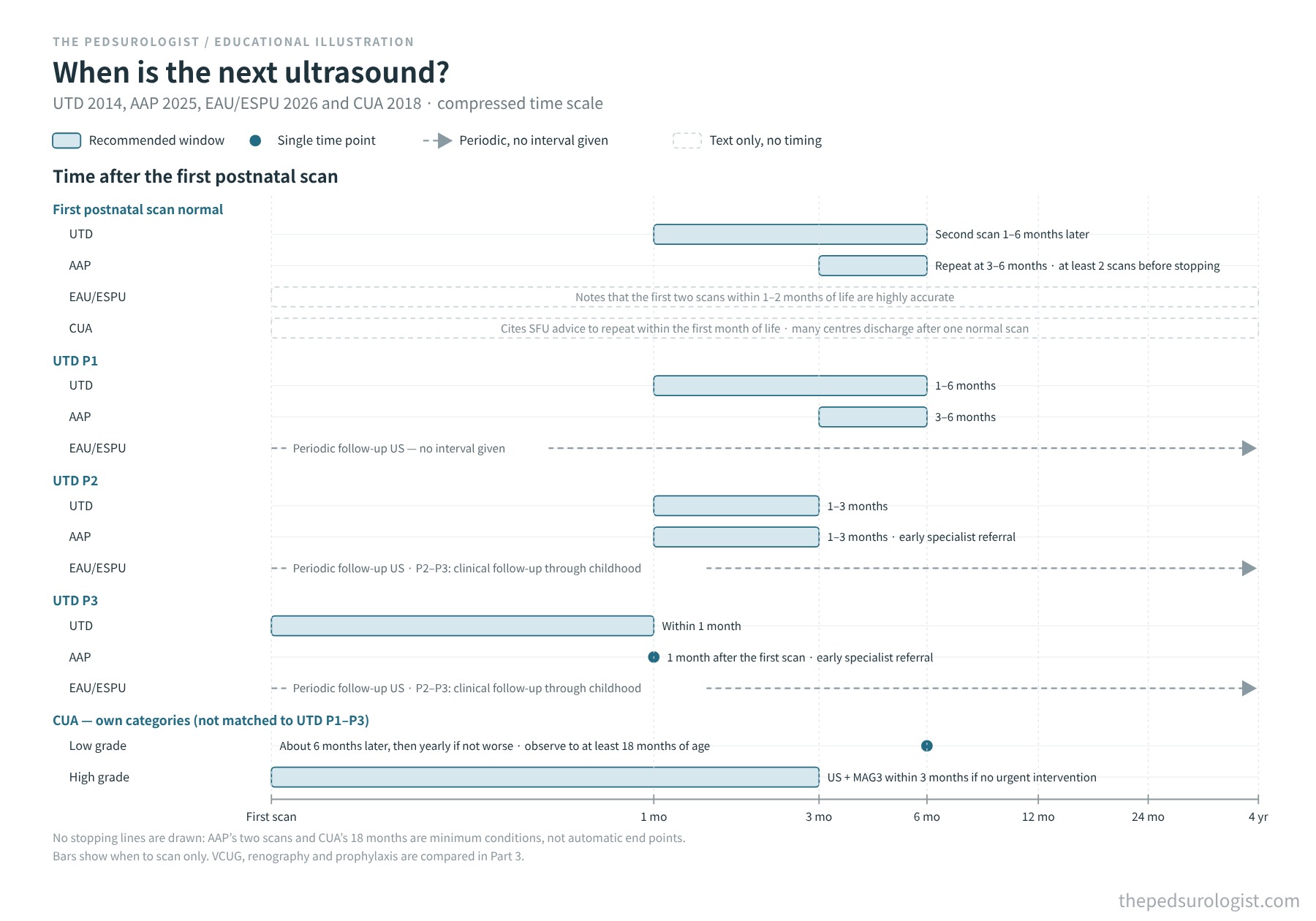
UTD and the AAP almost coincide in shortening the interval as risk rises: 1–6 and 3–6 months in P1, 1–3 months for both in P2, and within 1 month and at 1 month in P3. EAU/ESPU calls for periodic follow-up without an interval, and CUA gives 6 months and 3 months for categories defined differently. The documents part ways on whether to scan again after a normal first scan, and on how long to follow dilation that remains. Stopping is the weakest point in all four: the AAP’s two scans and CUA’s 18 months are only minimum conditions, and none defines how long persisting low-risk dilation should be followed.
Part 3 uses the same grade-by-grade structure to compare who should have VCUG and diuretic renography, and who should receive antibiotic prophylaxis.
Currency and scope of the evidence
Guideline texts checked: AAP 2025 on 30 October 2026 (full text, key recommendations; flowchart Figures 5 and 6 not compared), and EAU/ESPU 2026 (limited update March 2026) on 30 October 2026 (PDF, including Figure 10 of chapter 16, §17.2.2 of chapter 17 and chapter 28). The CUA/PUC document used here is the 2018 edition, which is listed in the CUAJ guideline index.
References
- Nguyen HT, Benson CB, Bromley B, et al. Multidisciplinary consensus on the classification of prenatal and postnatal urinary tract dilation (UTD classification system). J Pediatr Urol. 2014;10(6):982-998. doi:10.1016/j.jpurol.2014.10.002
- Herndon CDA, Otero HJ, Hains D, Sweeney RM, Lockwood GM; Section on Urology; Section on Nephrology; Section on Radiology; Section on Hospital Medicine. Perinatal urinary tract dilation: recommendations on pre-/postnatal imaging, prophylactic antibiotics, and follow-up: clinical report. Pediatrics. 2025;156(1):e2025071814. doi:10.1542/peds.2025-071814
- EAU Guidelines on Paediatric Urology, 2026 edition (limited update March 2026). Chapter 16, Dilatation of the upper urinary tract (PUJ obstruction), Figure 10; Chapter 17, Vesicoureteric reflux, §17.2.2; Chapter 28, Foetal urology, Figure 17 and Tables 14–15. European Association of Urology.
- Capolicchio JP, Braga LH, Szymanski KM. Canadian Urological Association/Pediatric Urologists of Canada guideline on the investigation and management of antenatally detected hydronephrosis. Can Urol Assoc J. 2018;12(4):85-92. doi:10.5489/cuaj.5094
- Nguyen HT, Herndon CDA, Cooper C, et al. The Society for Fetal Urology consensus statement on the evaluation and management of antenatal hydronephrosis. J Pediatr Urol. 2010;6(3):212-231. doi:10.1016/j.jpurol.2010.02.205