
When a prenatal scan shows renal pelvic dilation, the first decision is the ultrasound schedule. This part covers its first half: when to rescan before birth and when to perform the first postnatal ultrasound, compared by degree of risk across four documents. They are the UTD multidisciplinary consensus (Nguyen et al., 2014), the American Academy of Pediatrics (AAP) clinical report (Herndon et al., 2025), the EAU/ESPU paediatric urology guideline (2026) and the Canadian CUA/PUC guideline (Capolicchio et al., 2018). Follow-up intervals after the first scan, and when to stop, are in Part 2; VCUG, diuretic renography and antibiotic prophylaxis are in Part 3.
For the same degree of prenatal dilation, do the four documents recommend the first scan at the same time? If not, is it because they draw the grade boundaries differently, or because they judge the same grade differently?
1. How the four documents’ grades correspond
A grade-by-grade comparison first needs a map of how the four documents’ grades line up. The AAP opens its key recommendations by saying that dilation should be classified with the UTD system. The UTD consensus created that system, yet it says the underlying literature is inconsistent and limited and offers its management scheme as a starting point for observation and study. EAU/ESPU considers that no system has been shown to be superior or universally adopted, and does not recommend one classification over another. CUA combines SFU grade (Fernbach et al., 1993) with APD into low and high grade, and says the added complexity of UTD calls for more validation. The Onen grade (Onen, 2007) is not the reference system in any of the four.
Scroll sideways to see the whole figure
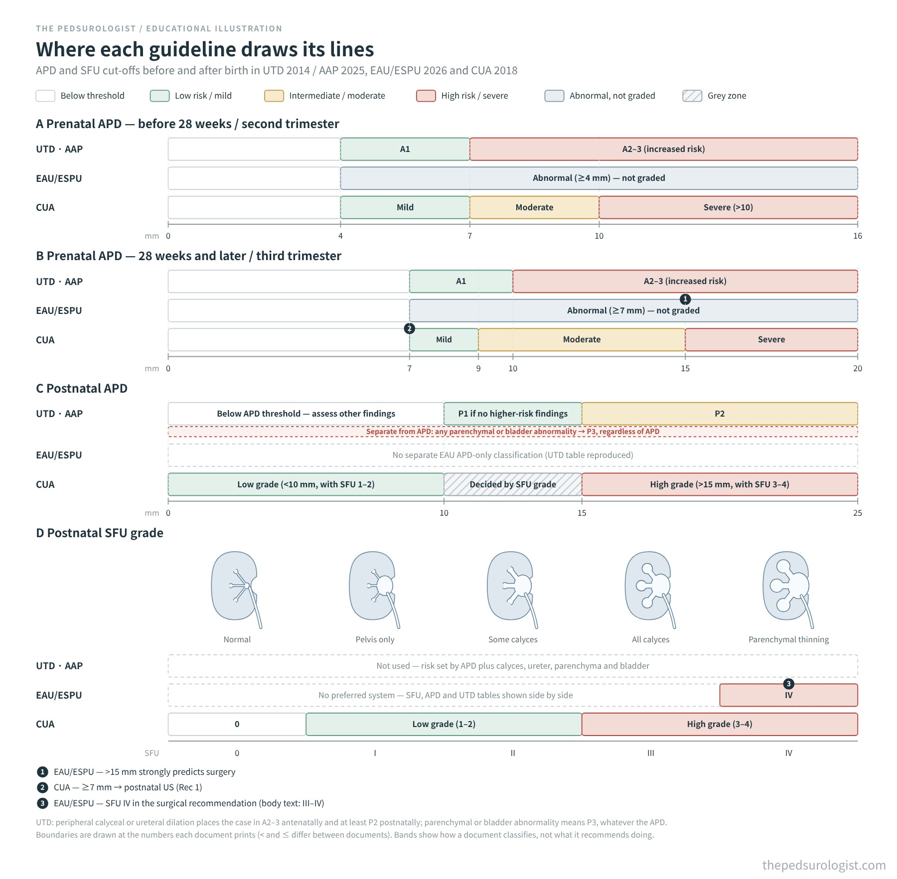
Reading Figure 1. Green is low risk or mild, yellow intermediate or moderate, red high risk or severe; grey marks a range called abnormal without a grade, hatching the range CUA assigns by SFU grade, and a dashed box a document that sets no threshold on that axis. ① EAU/ESPU: an antenatal APD above 15 mm strongly predicts postnatal surgery. ② CUA: a third-trimester APD of 7 mm or more indicates a postnatal ultrasound. ③ EAU/ESPU: its surgical recommendation uses SFU grade 4 (the body text says 3–4). UTD risk groups are not set by APD alone. Peripheral calyceal or ureteral dilation means A2–3 antenatally and at least P2 postnatally, and a parenchymal or bladder abnormality means P3 regardless of APD (the dashed red box under the UTD row in C). Central calyceal dilation makes a kidney P1 even with an APD below 10 mm, and an APD in the P1 range counts as P1 only if no higher-risk finding is present. Boundaries are drawn at the numbers each document prints; the use of “below” and “up to” differs between documents.
EAU/ESPU (the dashed boxes in Figure 1) reproduces both the SFU and the UTD tables. It gives numbers for the antenatal thresholds (4 mm before and 7 mm after 28 weeks) and states that an antenatal APD above 15 mm strongly predicts postnatal surgery (level of evidence 1b), and its text cites resolution, surgery and long-term outcome rates for UTD P1, P2 and P3. It sets no repeat-scan interval for each UTD group, but it does give timings for the first postnatal ultrasound in Figure 10 of the upper tract dilatation chapter, Figure 17 of the foetal urology chapter and the text of the reflux chapter. These three sources divide patients differently, their timings do not fully agree, and none maps one-to-one onto UTD A1 and A2–3, so the tables below list each EAU/ESPU source separately. Figure 17 adapts the pathway of Rickard et al. (2022); it is a management flowchart, and its boxes are not separate Strong recommendations.
As the figure shows, the correspondence between documents is approximate. A postnatal APD of at least 10 mm and below 15 mm with no higher-risk finding is UTD P1. In CUA the same APD range is a grey zone where SFU grade sets the pathway, and CUA’s high grade puts SFU 3 and 4 together, so it is not the same group as UTD P2 or P3. Whatever system is used, recording the APD value, central versus peripheral calyceal dilation, parenchymal thickness and echogenicity, ureteral diameter and bladder findings separately from the grade lets the case be read again against another document’s criteria. Onen grade 2 does not make a kidney UTD P2 (clinical Part 1). This part and Part 2 are organised by UTD group, with CUA’s own categories named in each table; CUA’s postnatal follow-up pathway is treated separately in Part 2.
The documents also differ in how they label recommendations. The UTD consensus and the AAP report carry no evidence grades; EAU/ESPU uses Strong and Weak, but its scan timings sit mostly in ungraded text and flowcharts; CUA grades only the five recommendations in its summary (level of evidence 3–4, grade C–D). The data behind all four are largely the same observational studies and expert consensus. A grade means something only within each document’s own method, and it is not a scale for comparing documents.
The American Urological Association (AUA) and the Society for Pediatric Urology (SPU) have no stand-alone hydronephrosis guideline. The SPU was one of the eight societies in the UTD consensus, and the AUA sponsored that meeting. The AUA’s recommendations on prenatal hydronephrosis sit within its reflux guideline and cover only screening for reflux, and NICE NG224 deals with evaluation after urinary tract infection, so neither is compared in this series. Reflux screening in infants with prenatal hydronephrosis is covered in VUR guidelines, Part 1 and will be linked again in the VCUG section of Part 3. The Indian Society of Pediatric Nephrology (ISPN) 2013 guideline predates UTD and is left out of this comparison.
2. Antenatal low risk (A1)
| Document | Prenatal follow-up | If it resolves before birth | If it persists: first postnatal ultrasound |
|---|---|---|---|
| UTD | If diagnosed before 32 weeks, rescan at 32 weeks (“at 32 weeks” in the original) | Resolved on the 32-week scan: no postnatal evaluation or follow-up | After 48 hours and before 1 month; a second scan 1–6 months later |
| AAP | If diagnosed before 32 weeks, one scan at 32 weeks or later | Resolution confirmed at 32 weeks or later: no postnatal evaluation. If the antenatal classification is incomplete or unclear, evaluate as A1 | Between 48 hours and 6 weeks of life. It can be ordered before discharge or at the first visit, and the birth admission need not be prolonged |
| EAU/ESPU | 32-week scan if there are red flags or the second-trimester APD is 4 mm or more or SFU 2 or more (Figure 17) | Resolved, or APD 7 mm or less or SFU 1 or less, with no red flags at 32 weeks: no follow-up (Figure 17) | Unilateral isolated dilation: days 7–14 in Figure 10; within 1 month in Figure 17 (32-week APD below 10 mm or SFU 2 or less). The reflux chapter text says to delay the scan until the first week after birth |
| CUA | At the obstetrician’s discretion | Not stated. A postnatal ultrasound is indicated for a third-trimester APD of 7 mm or more (Recommendation 1) | Low grade expected: physician’s discretion (1–3 months in the flowchart); avoid the first 2 days |
The 32-week point exists because many cases resolve before then. The AAP notes that about 80% of second-trimester APDs of 4–8 mm resolve before birth, and EAU/ESPU regards most antenatal dilation (up to 80%) as transient. A pathway that ends follow-up when dilation has resolved at 32 weeks with no red flags appears in UTD, the AAP report and EAU/ESPU Figure 17; CUA has no such sentence. Because the documents define red flags and resolution slightly differently, skipping postnatal imaging after antenatal resolution should take into account the gestational age at which resolution was seen, the earlier risk group and any associated findings. The data the AAP cites and the data EAU/ESPU quotes (44% of antenatally resolved cases reappeared after birth, but none needed surgery) both point to a very low risk of urinary tract infection, loss of function or surgery in these children.
When dilation persists, the recommended time for the first postnatal scan differs. UTD says before 1 month, the AAP within 6 weeks, and CUA leaves it to the physician (1–3 months in its flowchart). EAU/ESPU differs within itself: for unilateral isolated dilation Figure 10 gives days 7–14, Figure 17 gives within 1 month, and the reflux chapter says to delay until the first week after birth. The basis of the 48-hour rule is that physiological fluid shifts just after birth can make dilation measure smaller, and the AAP notes that this shift does not fully resolve until days 7–10. The AAP’s 6 weeks allows the scan to be ordered either before discharge or at the first visit, which reads as a way of planning so that outpatient imaging and follow-up are not missed. That reading is the author’s interpretation. The opening paragraph of the AAP key recommendations is about sharing responsibility so that tests and care needed after discharge are not lost.
3. Antenatal increased risk (A2–3)
| Document | Prenatal follow-up | Prenatal consultation and referral | First postnatal ultrasound |
|---|---|---|---|
| UTD | Rescan within 4–6 weeks of diagnosis; later intervals at discretion | Paediatric urology or nephrology consultation when the risk of surgery or renal dysfunction is substantial | After 48 hours and before 1 month; sooner for obstructive uropathy or bilateral disease |
| AAP | Every 4 weeks until delivery | Antenatal consultation for A2–3 and for bilateral dilation | If A2–3 at any time in pregnancy: during the birth admission, ideally after 48 hours. Without suspected outlet obstruction the admission is not prolonged for this scan |
| EAU/ESPU | 32-week scan (Figure 17) | Multidisciplinary care for urinary tract malformations (Strong) | Unilateral isolated dilation with a 32-week APD of 10 mm or more or SFU 3–4: 48 hours to 1 week; bilateral isolated dilation: after 48 hours (Figure 17). With ureteral dilation, from within 24 hours to within 1 week depending on sex and laterality |
| CUA | At the obstetrician’s discretion | Severe bilateral dilation or oligohydramnios is often referred for antenatal counselling | High grade expected: as soon as feasible after day 2, usually within 2 weeks; sooner with ureteral dilation, bladder abnormality or parenchymal abnormality |
The higher the risk, the earlier the first scan, though the windows differ in width: during the birth admission for the AAP, 48 hours to under 1 week on some EAU/ESPU Figure 17 pathways, usually within 2 weeks for CUA, and before 1 month for UTD. The targets for antenatal consultation are similar. That CUA sets no prenatal interval is not an objection; it leaves prenatal follow-up to obstetrics.
4. Severe bilateral dilation and suspected outlet obstruction
| Document | When not to wait 48 hours | What else to do |
|---|---|---|
| UTD | Oligohydramnios, urethral obstruction, bilateral high-grade dilation | — |
| AAP | Suspected bladder outlet obstruction (posterior urethral valves and others). The report’s recommendations are not primarily aimed at severe bilateral dilation | Within 48 hours of life: urology and nephrology consultation, bladder decompression, imaging and antibiotic prophylaxis. Serum creatinine in the first days reflects the maternal value |
| EAU/ESPU | Severe cases (bilateral dilation, solitary kidney, oligohydramnios): immediately (text); 0–48 hours in Figure 10. Bladder abnormality or reduced amniotic fluid: within 24 hours in Figure 17 | Figure 10: further studies to exclude hydroureteronephrosis, posterior urethral valves, neurogenic bladder and others |
| CUA | When immediate management is needed, as with posterior urethral valves | Urgent VCUG if outlet obstruction is suspected. Serum creatinine after day 2 in severe bilateral dilation, abnormal renal echogenicity or a solitary kidney |
The exceptions are almost the same in all four documents. If outlet obstruction is suspected, bilateral dilation is severe or there was oligohydramnios, the 48 hours are not awaited. Bilateral dilation alone is not a reason to scan everyone at once: EAU/ESPU Figure 17 also separates isolated bilateral dilation (after 48 hours) from bladder abnormality (within 24 hours).
Closing Part 1
Putting the first-scan timings from sections 2–4 on one page gives Figure 2.
Scroll sideways to see the whole figure
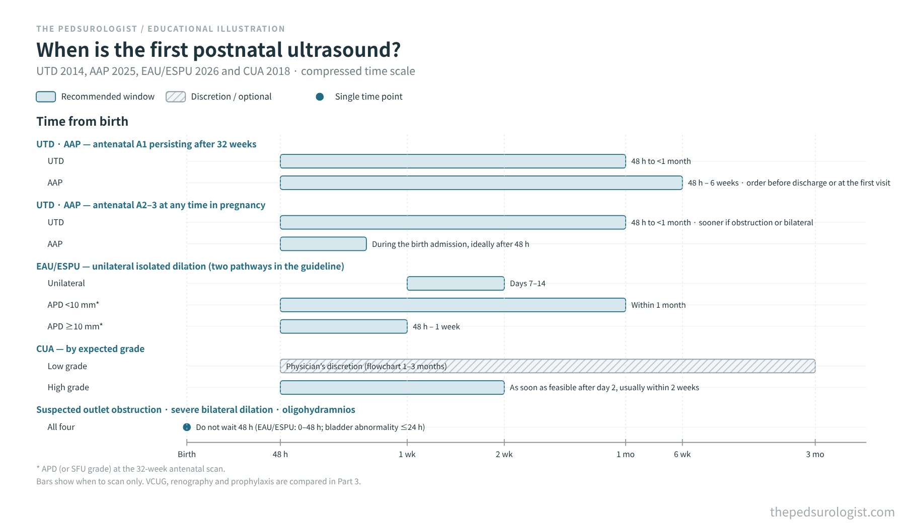
Seen this way, the differences between the documents have two sources. One is where the lines are drawn: when a document divides categories differently from UTD, as CUA does, the same child can be given a different schedule. The other is judgement: whether a low-risk child’s first scan should be at 1–2 weeks or left open until 6 weeks depends on what weight is given to the same data. EAU/ESPU’s flowcharts and text even disagree with each other. The exceptions to the 48-hour rule, by contrast, are almost identical across the four.
Whichever document is followed, recording the individual findings lets the case be read again against another document’s criteria, and the absence of an item from one document does not mean that document opposes it. Part 2 compares, after the first scan, when the next ultrasound is performed and when follow-up ends, for a normal first scan and for P1, P2 and P3.
Currency and scope of the evidence
Guideline texts checked: AAP 2025 on 30 October 2026 (full text, key recommendations; flowchart Figures 5 and 6 not compared), and EAU/ESPU 2026 (limited update March 2026) on 30 October 2026 (PDF, including Figure 10 of chapter 16, §17.2.2 of chapter 17 and Figure 17 of chapter 28). The CUA/PUC document used here is the 2018 edition, which is listed in the CUAJ guideline index. The British Association for Paediatric Nephrology (BAPN) published the scope for an antenatal hydronephrosis guideline in 2025.
References
- Nguyen HT, Benson CB, Bromley B, et al. Multidisciplinary consensus on the classification of prenatal and postnatal urinary tract dilation (UTD classification system). J Pediatr Urol. 2014;10(6):982-998. doi:10.1016/j.jpurol.2014.10.002
- Herndon CDA, Otero HJ, Hains D, Sweeney RM, Lockwood GM; Section on Urology; Section on Nephrology; Section on Radiology; Section on Hospital Medicine. Perinatal urinary tract dilation: recommendations on pre-/postnatal imaging, prophylactic antibiotics, and follow-up: clinical report. Pediatrics. 2025;156(1):e2025071814. doi:10.1542/peds.2025-071814
- EAU Guidelines on Paediatric Urology, 2026 edition (limited update March 2026). Chapter 16, Dilatation of the upper urinary tract (PUJ obstruction), Figure 10; Chapter 17, Vesicoureteric reflux, §17.2.2; Chapter 28, Foetal urology, Figure 17 and Tables 14–15. European Association of Urology.
- Capolicchio JP, Braga LH, Szymanski KM. Canadian Urological Association/Pediatric Urologists of Canada guideline on the investigation and management of antenatally detected hydronephrosis. Can Urol Assoc J. 2018;12(4):85-92. doi:10.5489/cuaj.5094
- Fernbach SK, Maizels M, Conway JJ. Ultrasound grading of hydronephrosis: introduction to the system used by the Society for Fetal Urology. Pediatr Radiol. 1993;23(6):478-480. doi:10.1007/BF02012459
- Onen A. An alternative grading system to refine the criteria for severity of hydronephrosis and optimal treatment guidelines in neonates with primary UPJ-type hydronephrosis. J Pediatr Urol. 2007;3(3):200-205. doi:10.1016/j.jpurol.2006.08.002
- Rickard M, Dos Santos J, Keunen J, Lorenzo AJ. Prenatal hydronephrosis: bridging pre- and postnatal management. Prenat Diagn. 2022;42(9):1081-1093. doi:10.1002/pd.6114