
"The ultrasound during pregnancy showed that our baby's kidney is swollen. They called it hydronephrosis. Will our baby need surgery after birth?"
Parents who hear the word hydronephrosis on a prenatal ultrasound often spend the weeks or months left before delivery feeling anxious. There are two things I want to say first. First, hydronephrosis is a common finding on prenatal ultrasound (about 1–5 in 100 pregnancies, depending on the criteria used and the stage of pregnancy)[8]. Second, mild dilation usually improves as the baby grows.[1] Still, not every baby follows the same course, so a few things need to be checked after birth. This article explains what hydronephrosis is, what the grades mean, and which tests your baby will have after birth and when, in the order we go through them in clinic.
Hydronephrosis is a finding, not a diagnosis
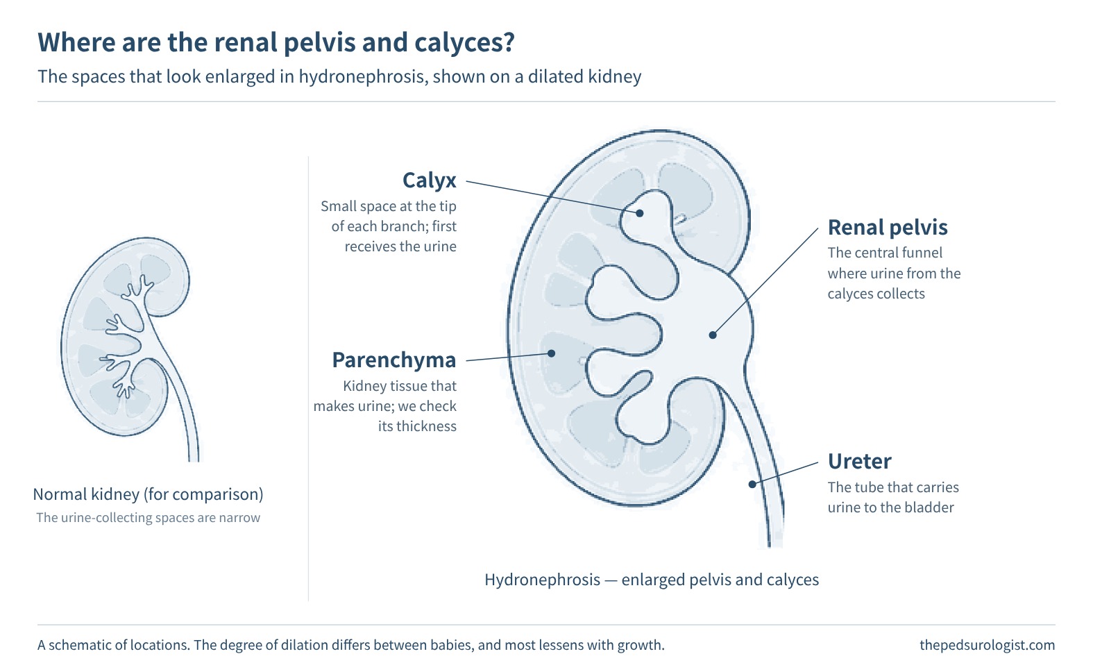
Urine made in the kidney passes through small spaces inside the kidney (the calyces) and collects in a funnel-shaped space in the middle (the renal pelvis). From there it travels down the ureter to the bladder. Hydronephrosis means these spaces look enlarged. So hydronephrosis is not the name of a disease. It is a word for what is seen on ultrasound, and the word alone does not tell you the cause. Several different causes can lie behind it.
Scroll horizontally to see the full figure
- Temporary dilation — the most common. One explanation is that the movements a baby's ureter uses to push urine along are still immature. The dilation often shrinks or disappears as the baby grows. But the first scan alone cannot confirm that it is temporary, so the course is followed over time.
- Ureteropelvic junction obstruction — urine does not drain well where the renal pelvis meets the ureter. This is the most common of the disease-related causes.[7]
- Vesicoureteral reflux — urine in the bladder flows backward up toward the kidney.
- Dilation that extends into the ureter (megaureter) — this can happen when urine does not drain well at the lower end of the ureter or when there is reflux. Sometimes the ureter is enlarged without any clear blockage.
- Rarely, the cause may be valves in the urethra, or a duplex kidney, in which the urine-collecting space and the ureter of one kidney are split into two. A kidney with many cysts (multicystic dysplastic kidney) can look like hydronephrosis, so the two need to be told apart.
Scroll horizontally to see the full figure
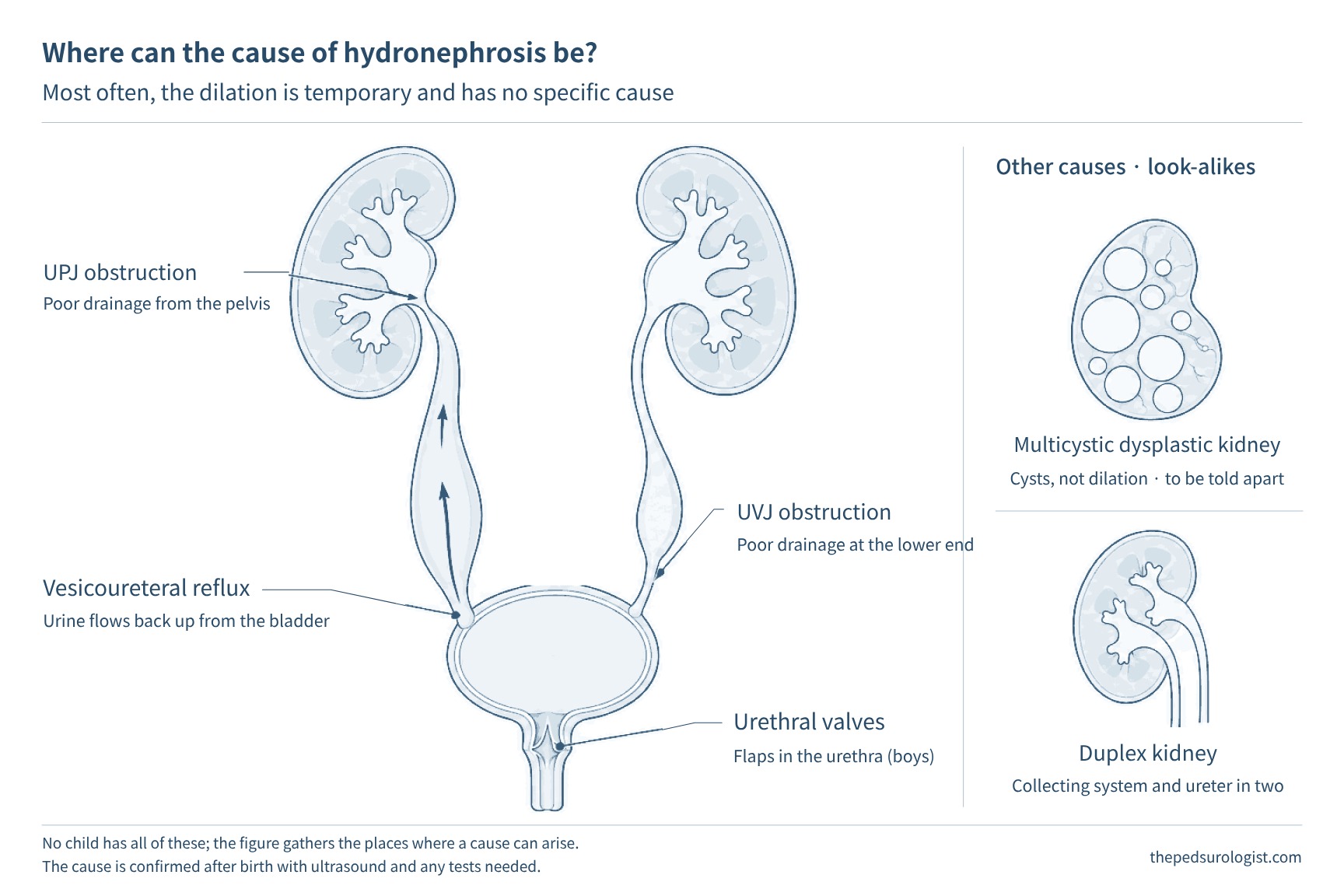
A prenatal ultrasound can suggest the cause, but it is often hard to be sure. That is why tests after birth are needed.
What do the grades and numbers mean?
The ultrasound report may include a number such as "renal pelvis 8 mm" or a grade such as "grade 2" or "UTD A2–3". The number is the front-to-back width of the renal pelvis (the anteroposterior renal pelvic diameter). The grade describes how much the renal pelvis and calyces are enlarged and whether the kidney tissue has become thin; the UTD classification also includes the ureter and bladder. Hospitals use different grading systems, so the same baby may be described differently.[2]
Scroll horizontally to see the full figure
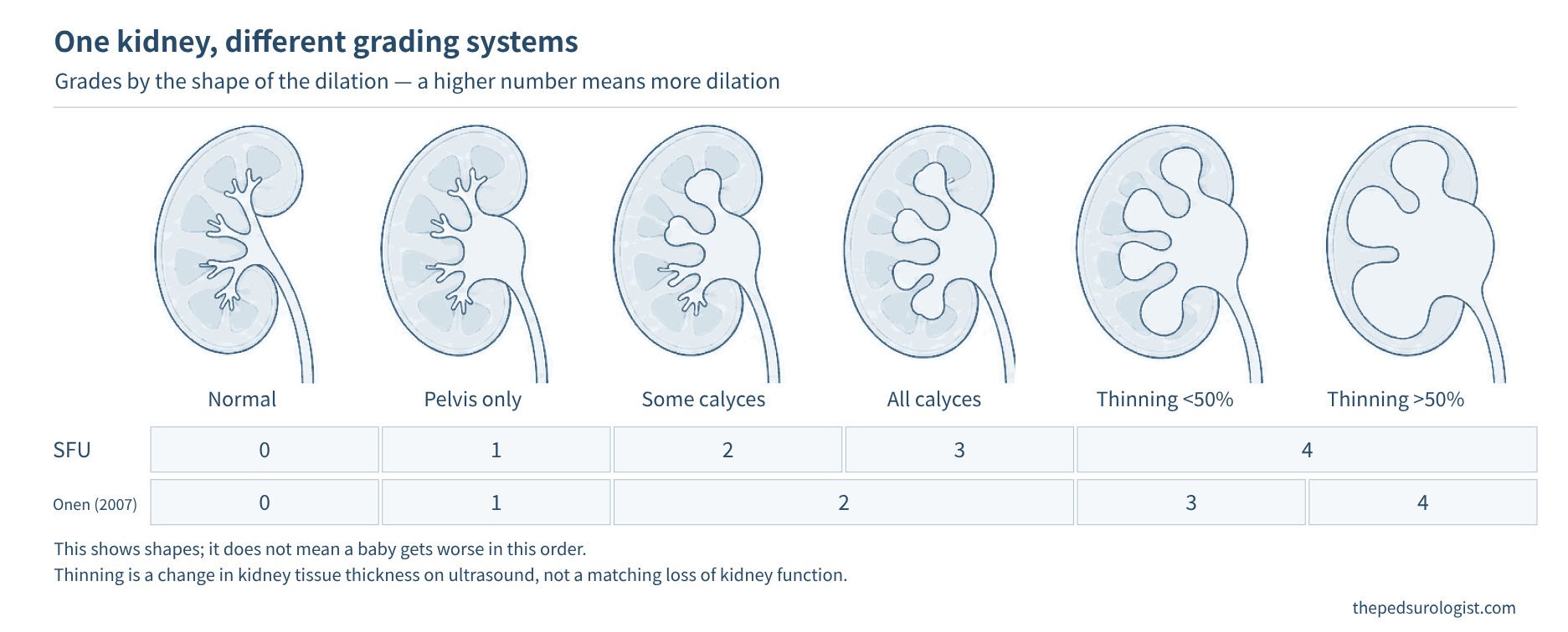
The grade tells you how marked the dilation is and how closely it needs to be watched. The higher the grade, the more likely it is that an underlying condition will be found after birth, and the longer it may take to improve or the longer the dilation may last.[3] Even so, a high grade does not automatically mean surgery. At the milder end, a large registry study found that about 90% of babies classified as low risk after birth had returned to normal by age 4.[1] Studies differ in who they included and in how they defined "improved," so no single number can be applied directly to your baby. Your baby's course is best judged from your baby's own tests.
Just as important as the grade are the ureter, the bladder and the kidney tissue. Compared with a renal pelvis that is only slightly enlarged, testing and management become more active when the ureter is also enlarged, the bladder looks abnormal, or the kidney tissue looks thin. If your report includes phrases such as "ureteral dilation," "bladder abnormality" or "parenchymal thinning," be sure to go over them at your visit.
Which tests after birth, and when?
The first ultrasound is usually done after the baby is 48 hours old. For the first two days or so, a baby makes little urine, so a scan done too early can make the hydronephrosis look milder than it really is. If the dilation was mild, it is usually done around one month of age; if it was large before birth, it is done earlier, while the baby is still in the hospital or soon after going home. The exact timing varies a little between guidelines and hospitals, so it helps to settle it before delivery with the hospital where your baby will be seen.[1][2]
However, if both kidneys are severely enlarged, if a baby's only kidney is enlarged, if there was little amniotic fluid, or if a blockage of the urethra is suspected (especially in boys), the baby is assessed right after birth.
For babies who need tests after birth, the ultrasound is repeated once at 3–6 months, even if the first one is normal. This is because dilation may not show well in the early newborn period.[1] The exception is a baby who had only mild dilation before birth that had already returned to normal on an ultrasound after 32 weeks of pregnancy. In that case, tests after birth are considered unnecessary.[1][2] If the dilation was ever large before birth, or if the prenatal records are unclear, the baby has tests after birth.
Further tests depend on the ultrasound results.
| Test | What it shows | When it is usually done |
|---|---|---|
| Follow-up ultrasound | Whether the dilation is shrinking or growing, whether the kidney tissue is preserved, and whether the kidney is growing well | Babies who need follow-up after birth. The interval ranges from 1 to 6 months, depending on risk |
| Reflux test (VCUG) | Whether urine in the bladder flows backward, and whether there is a problem with the urethra or bladder | When the ureter is markedly enlarged, when the kidney tissue or bladder is abnormal, or when the baby has had a urinary tract infection with fever. For moderate dilation, it is decided for each baby after discussion |
| Renal scan (diuretic renogram) | How the work is shared between the two kidneys, and whether urine drains well | When the kidney tissue looks thin, or when the dilation is severe or getting larger, among other reasons. Timing is set for after about 1–3 months of age, once the kidneys have matured somewhat |
Most babies with mild dilation and normal kidney tissue, ureters and bladder are followed with repeat ultrasounds only. For the reflux test, a thin tube is passed through the urethra and the bladder is filled with contrast dye. The test then checks whether the dye flows backward toward the kidney while the baby passes urine. That is why it is done only in babies who really need it.[6]
Does my baby need antibiotics?
Preventive (prophylactic) antibiotics are a small dose of medicine given every day to lower the risk of a urinary tract infection before one happens. There was a time when almost every baby with hydronephrosis was given them, but that is no longer the case. In babies whose ureters and bladder are normal and whose dilation is limited to the renal pelvis, urinary tract infections are uncommon to begin with (about 4%), and infection rates did not differ between babies who received antibiotics and babies who did not.[4] So with mild dilation, they are usually not given.
On the other hand, for high-risk babies whose ureter is markedly enlarged (7 mm or more) or whose kidney tissue or bladder is abnormal, urinary tract infections are more likely, so preventive antibiotics are recommended. When vesicoureteral reflux is confirmed, the decision also takes into account the grade of reflux and any history of urinary tract infection.[1][5] Girls and boys who have not had a circumcision are also at higher risk of infection, but the decision is not based on sex or circumcision alone. It is weighed together with the ultrasound findings. If antibiotics are started, how long to give them and when to reassess are discussed too. Most urinary tract infections with fever happen in the first year of life, so around the first birthday, reflux, the degree of ureteral dilation and the course of any infections are reviewed again to decide whether to continue the antibiotics. Do not stop prescribed antibiotics on your own, and do not give leftover medicine later.
What to watch for at home, and when to seek care right away
The most important thing for a baby with hydronephrosis is not to miss a urinary tract infection when there is a fever. A baby cannot tell you that it hurts to pass urine, and a urinary tract infection can be present even when there are cold symptoms such as a runny nose or cough. When your baby is seen for a fever, be sure to say that hydronephrosis was found before birth, and ask whether a urine test is needed.
If a baby younger than 3 months has a fever of 38°C or higher, or has a fever along with poor feeding, listlessness or vomiting, seek care right away. Do not wait either if your baby is passing noticeably less urine or the belly looks swollen.
Going to follow-up ultrasounds on the scheduled dates also matters. In particular, for a baby whose hydronephrosis was severe at first, it is safer to keep watching for a while even after it improves. This is because some children who improved in infancy come back years later with flank pain or a urinary tract infection with fever. If these symptoms appear even after follow-up has ended, see a doctor again.
Frequently asked questions
Is there anything we can do during pregnancy?
Most hydronephrosis needs no treatment during pregnancy, and there is rarely any need to change how or when the baby is delivered. The course is followed with prenatal ultrasounds. If the dilation is large or is severe on both sides, talking with a pediatric urologist or pediatric nephrologist before delivery makes it easier to plan for after birth.
When would surgery be done?
Only some babies need surgery. Surgery suited to the cause is considered when, during follow-up, the dilation keeps getting larger, a renal scan shows that one kidney's function is falling, or urinary tract infections with fever or pain keep coming back. Usually the decision is based on change over time rather than on a single test. But if a serious problem is found on the first tests, treatment may begin right away without waiting for repeated tests.
We were told it has improved. Do we still need to come back?
If the dilation was mild at first and has been confirmed normal on two or more ultrasounds, follow-up can end. But if it was severe at first, it is a good idea to check a few more times even after it improves. Talk with your child's doctor about how long follow-up should continue.
There are articles covering the grading systems for hydronephrosis (APD, SFU, Onen, UTD), its natural history, postnatal evaluation, and the evidence on prophylactic antibiotics.
Hydronephrosis, Part 1 — What We Measure and How We Grade It
Hydronephrosis, Part 2 — Who Needs What, and When, After Birth
Hydronephrosis, Part 3 — How the Ureter Forms, and Why It Dilates
Content for healthcare professionals.
References
- Herndon CDA, Otero HJ, Hains D, et al. Perinatal urinary tract dilation: recommendations on pre-/postnatal imaging, prophylactic antibiotics, and follow-up: clinical report. Pediatrics. 2025;156(1):e2025071814. doi:10.1542/peds.2025-071814
- Nguyen HT, Benson CB, Bromley B, et al. Multidisciplinary consensus on the classification of prenatal and postnatal urinary tract dilation (UTD classification system). J Pediatr Urol. 2014;10(6):982-998. doi:10.1016/j.jpurol.2014.10.002
- Lee RS, Cendron M, Kinnamon DD, Nguyen HT. Antenatal hydronephrosis as a predictor of postnatal outcome: a meta-analysis. Pediatrics. 2006;118(2):586-593. doi:10.1542/peds.2006-0120
- Chamberlin JD, Braga LH, Davis-Dao CA, et al. Continuous antibiotic prophylaxis in isolated prenatal hydronephrosis. J Pediatr Urol. 2022;18(3):363.e1-363.e7. doi:10.1016/j.jpurol.2022.03.027
- Holzman SA, Braga LH, Zee RS, et al. Risk of urinary tract infection in patients with hydroureter: an analysis from the Society of Fetal Urology Prenatal Hydronephrosis Registry. J Pediatr Urol. 2021;17(6):775-781. doi:10.1016/j.jpurol.2021.09.001
- EAU Guidelines on Paediatric Urology. Dilatation of the upper urinary tract (PUJ obstruction); Foetal urology. European Association of Urology, 2026. uroweb.org
- Kim SW. Management of hydronephrosis: a comprehensive review in pediatric urology perspective. Child Kidney Dis. 2024;28(2):59-65. doi:10.3339/ckd.24.011
- Sinha A, Bagga A, Krishna A, et al. Revised guidelines on management of antenatal hydronephrosis. Indian Pediatr. 2013;50(2):215-231. doi:10.1007/s13312-013-0064-6